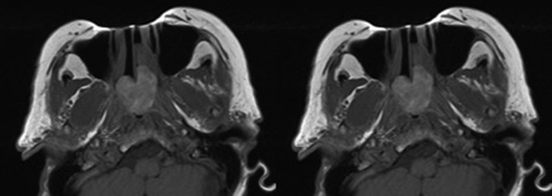
Fig. 2.

Preoperative T1-weighted post-contrast MRI images demonstrating a large polypoid mass originating from the left posterior nasal cavity protruding into the nasopharynx and filling the right choana (left). Postoperative scans with expected postsurgical changes but no evidence of disease (right). MRI, magnetic resonance imaging.
