
Fig. 2.
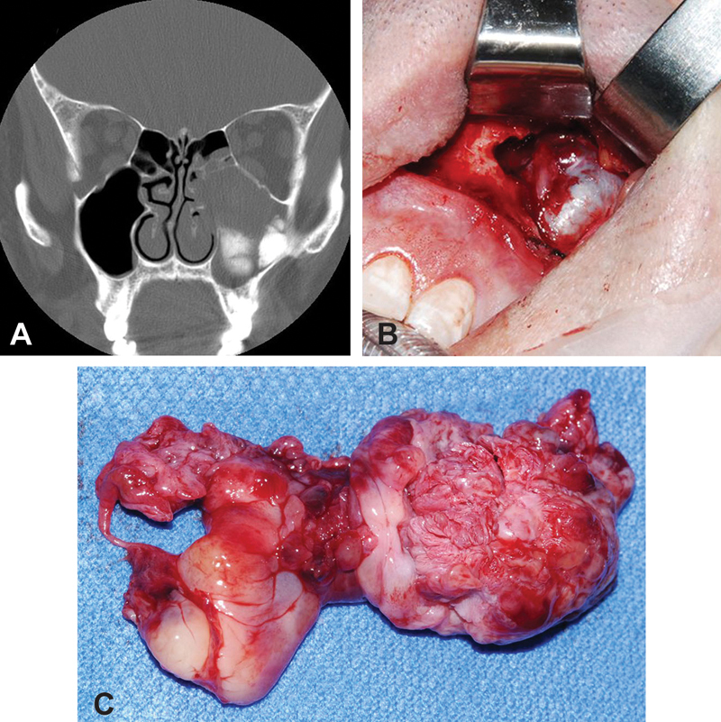
( A ) Coronal computed tomography of the paranasal sinuses demonstrates dense calcification inferiorly in an opacified and mildly expanded left maxillary sinus. ( B ) Intraoperative view of the left maxillary sinus with Caldwell–Luc approach and view of the tumor in situ ( C ) Specimen of osteosarcoma of the left maxillary sinus.