
Fig. 3.
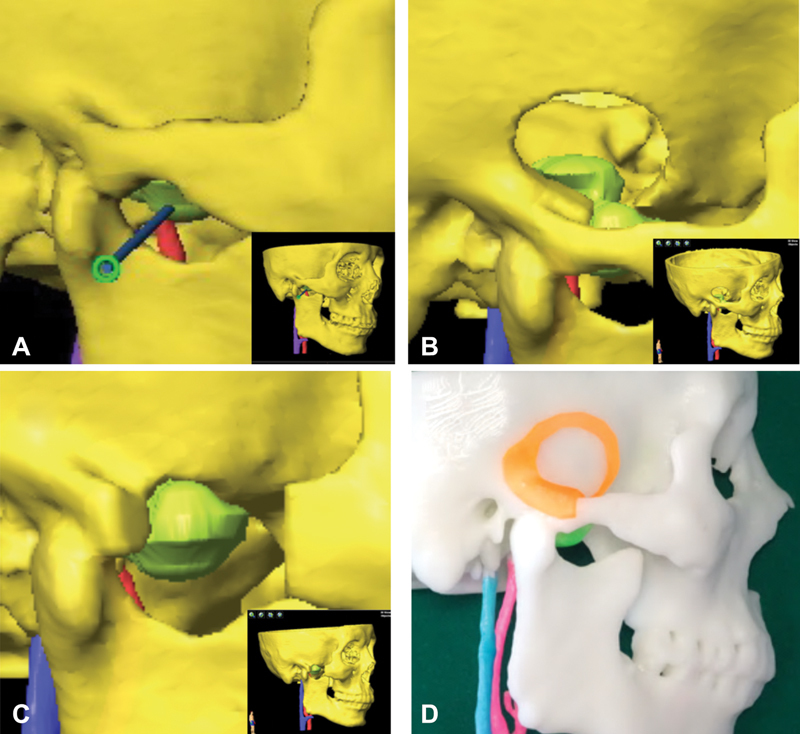
Three-dimensional (3D) model reconstruction and virtual planning. ( A ) The trajectory path design of navigation-guided core needle biopsy. ( B ) Lateral temporal craniotomy was designed for intracranial part removal of the lesion. ( C ) Zygomatic osteotomy was designed for extracranial part removal of the lesion. ( D ) 3D model with skull, tumor and vessels, and surgical guide plate (orange) were printed. Blue color column indicates the internal jugular vein (IJV), and red color column indicates the internal carotid artery (ICA). The inset shows the 3D image of the overall skull-tumor-vessel model derived from fusion imaging.