
Fig. 6.
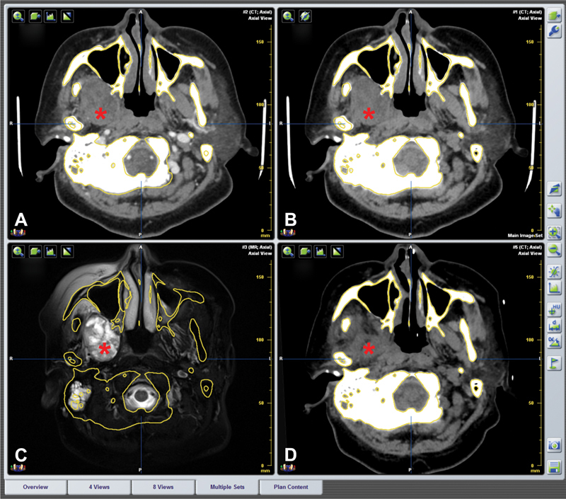
Fusion images of preoperation and postoperation in the same reference frame. ( A–C ) Preoperative contrast CT, plain CT, and MRI image. Asterisk indicates the tumor area. ( D ) No obvious tumor mass was found in postoperative CT image. The interspace was occupied by muscles and other soft tissues (asterisk).