
Figure 5.
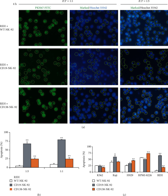
CD19-NK-92 and CD138-NK-92 cells selectively induced apoptosis of target cancer cells. Human ALL-derived REH cells were labeled with PKH67 dye; incubated with WT-NK-92, CD19-NK-92, or CD138-NK-92 cells at the E : T ratio of 1 : 1 or 1 : 5 for 4 h; stained with Hoechst 33342; and visualized under a fluorescence microscope. (a) Micrographs of PKH67-positive cells under green fluorescence were used to mark the target cells and apoptosis of target cells was scored under blue florescence based on the fragmented/condensed nuclei. (b) Percentage of apoptosis over total REH cells was plotted. Data are mean ± s.d. (n = 3). ∗p < 0.05 and ∗∗p < 0.01 versus WT-NK-92 cells at the same condition; two-sided Student's t-test. (c) Apoptosis of PKH67-labeled CML-derived K562, BL-derived Raji, MM-derived H929 and RPMI-8226, and ALL-derived REH cells upon incubation with WT-NK-92, CD19-NK-92, or CD138-NK-92 cells at the E : T ratio of 1 : 5 for 4 h was similarly determined by Hoechst 33342 assay. Percentage of apoptosis over total target cells was plotted. Data are means ± s.d. (n = 3). ∗p < 0.05 and ∗∗p < 0.01 versus WT-NK-92 cells; two-sided Student's t-test.