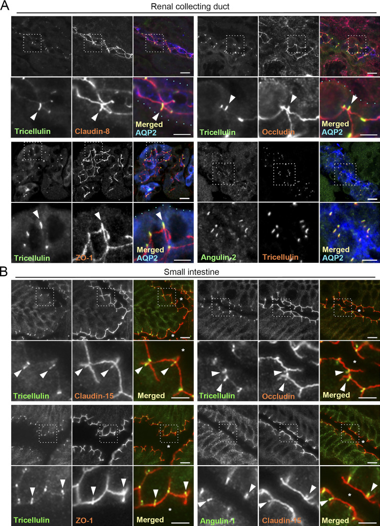
Figure 1.
Localization of tTJ and TJ proteins in the mouse kidney and small intestine. (A) Triple-immunofluorescence staining of frozen mouse kidney sections containing collecting ducts with anti-tricellulin mAb, anti–claudin-8 pAb, and anti–AQP-2 pAb (top left); anti-tricellulin mAb, anti-occludin pAb, and anti–AQP-2 pAb (top right); anti-tricellulin mAb, anti–ZO-1 pAb, and anti–AQP-2 pAb (bottom left); and anti-angulin-2 pAb, anti-tricellulin mAb, and anti–AQP-2 pAb (bottom right). AQP-2 staining is only shown in the merged images. The boxed regions are magnified on the bottom. Tricellulin shows rodlike staining in AQP-2–positive collecting ducts. Claudin-8, occludin, and ZO-1 colocalize with tricellulin at TCs (arrowheads). Light blue dots in the magnified merged images indicate the outline of collecting ducts. (B) Double-immunofluorescence staining of frozen mouse small intestine sections with anti-tricellulin mAb and anti–claudin-15 pAb (top left), anti-tricellulin mAb and anti-occludin pAb (top right), anti-tricellulin mAb and anti–ZO-1 pAb (bottom left), and anti–angulin-1 mAb and anti–claudin-15 pAb (bottom right). Asterisks show the intestinal lumen. The boxed regions are magnified on the bottom. Bars: 10 µm (top), 5 µm (bottom).

