Figure 2.
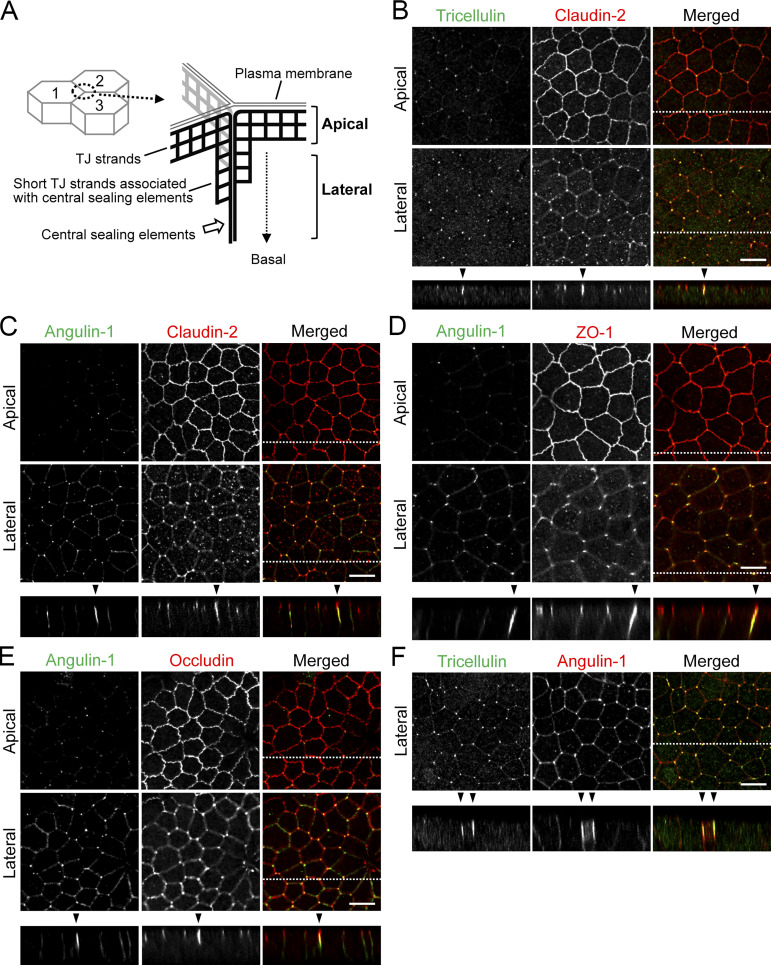
Localization of tTJ and TJ proteins at TCs in MDCK II cells. (A) Supposed relationship between tTJ structural organization and confocal sections in the apical and lateral regions. The scheme represents tTJs observed from the inside of cell 3. The network of black bold lines represents TJ strands. The most apical elements of TJ strands in bicellular TJs join together at TCs, turn, and extend in the basal direction attached to one another to form the central sealing elements that connect with short TJ strands. The confocal sections in the apical region correspond to the level of bicellular TJs, while those in the lateral region are more basal and do not contain bicellular TJs. (B–F) Double-immunofluorescence staining of MDCK II cells with anti-tricellulin pAb and anti–claudin-2 mAb (B), anti–angulin-1 pAb and anti–claudin-2 mAb (C), anti–angulin-1 pAb and anti–ZO-1 mAb (D), anti–angulin-1 pAb and anti-occludin mAb (E), and anti-tricellulin mAb and anti–angulin-1 pAb (F). Confocal sections in the apical region, including TJ markers and the lateral region, together with the corresponding Z-stack images along the white dotted lines are shown. Not only tricellulin and angulin-1 but also claudin-2, occludin, and ZO-1 show extended localization along the apicobasal axis at TCs (arrowheads). Bar: 10 µm.

