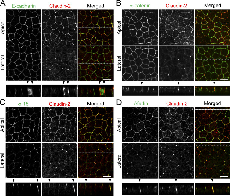
Figure 3.
Localization of tTJ and AJ proteins at TCs in MDCK II cells. (A–D) Double-immunofluorescence staining of MDCK II cells with anti–claudin-2 mAb and anti–E-cadherin mAb (A), anti–α-catenin pAb (B), anti–tension-dependent epitope of α-catenin mAb (α-18; C), and anti-afadin pAb (D). Confocal sections in the apical region, including claudin-2 signals at bicellular contacts and the lateral region, together with the corresponding Z-stack images along the white dotted lines are shown. α-18 under tension and afadin show extended localization along the apicobasal axis at TCs (arrowheads). Bar: 10 µm.

