Figure S5.
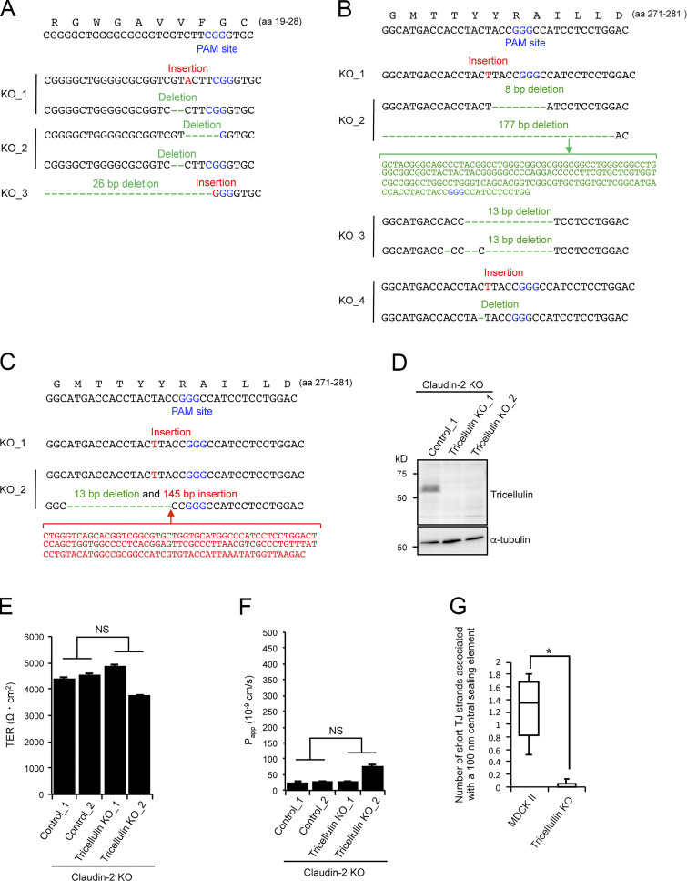
Characterization of claudin/angulin-1–KO cells, tricellulin-KO cells, and tricellulin/claudin-2–dKO cells. (A) Frameshift mutations introduced in the proximity of the PAM site in the angulin-1 gene in three claudin/angulin-1–KO clones (KO_1–3) established by CRISPR/Cas9-mediated genome editing. (B) Frameshift mutations introduced in the proximity of the PAM site in the tricellulin gene in four tricellulin-KO clones (KO_1–4) established by CRISPR/Cas9-mediated genome editing. (C) Frameshift mutations introduced in the proximity of the PAM site in the tricellulin gene in two tricellulin/claudin-2–dKO cell clones (KO_1 and KO_2) established by CRISPR/Cas9-mediated genome editing. (D) Western blotting of lysates of a control claudin-2–KO cell clone and two tricellulin/claudin-2–dKO cell clones with anti-tricellulin pAb or anti–α-tubulin mAb. (E and F) TER (E) and paracellular flux of fluorescein (F) were measured in two control claudin-2–KO cell clones (Control_1 and 2) and two tricellulin/claudin-2–dKO cell clones (KO_1 and KO_2). Data are shown as mean ± SD (n = 3) and were analyzed by Welch’s t test (n = 6 total for two control clones [n = 3 per clone] versus n = 6 total for two tricellulin/claudin-2–dKO clones [n = 3 per clone]). Papp, apparent permeability. (G) Quantification of short TJ strands associated with a central sealing element in MDCK II cells and tricellulin–KO_4 cells. Data were analyzed by Mann-Whitney U test (MDCK II cells, n = 17; tricellulin-KO cells, n = 16). *, P < 0.01.

