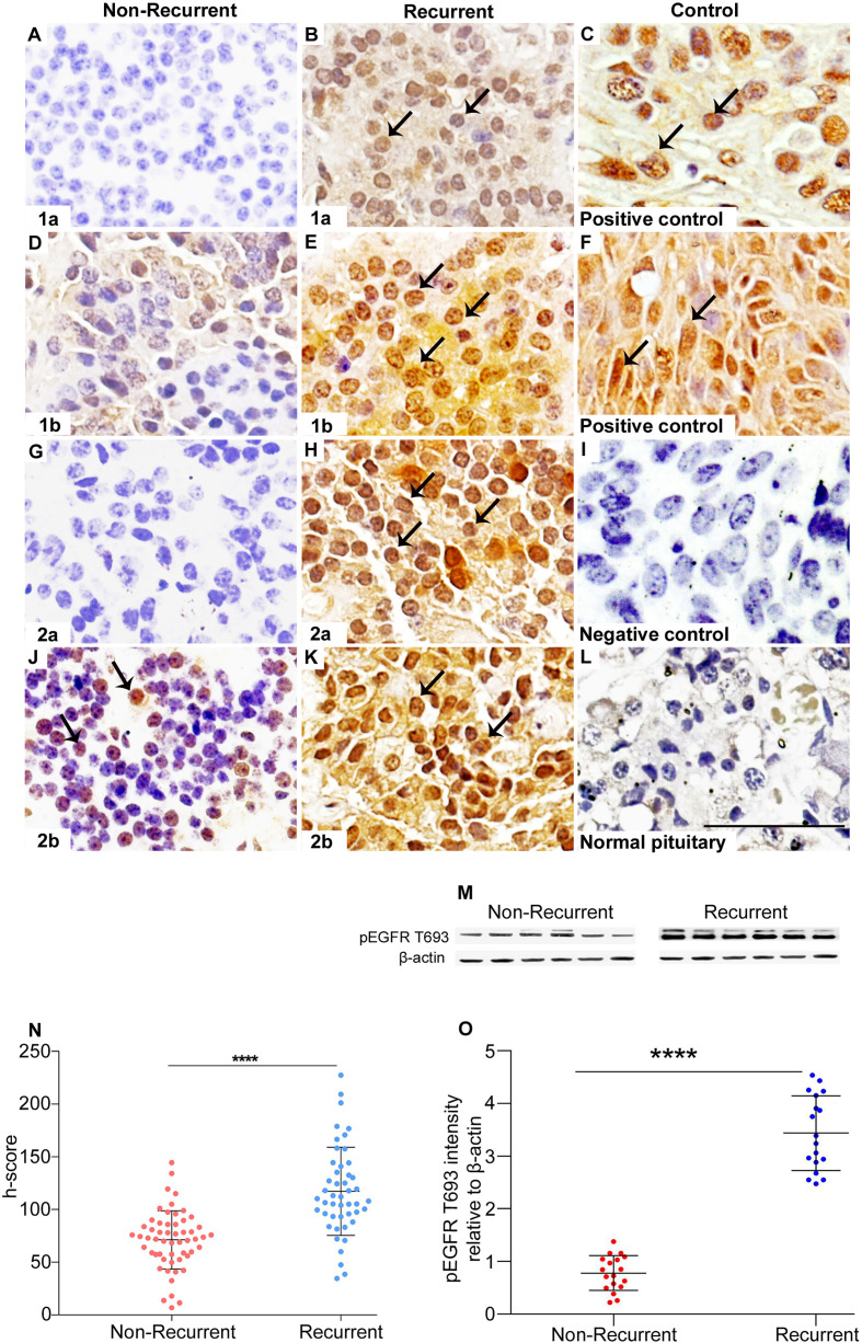
Figure 1.
Overexpression of pEGFR T693 in recurrent NFPAs. Immunohistochemistry of pEGFR T693 showed overexpression in recurrent NFPAs (B, E, H, K) as compared to non-recurrent NFPAs (A, D, G, J). pEGFR T693 showed strong nuclear positivity (brown) marked by black arrows. Breast cancer (C) and cervical cancer (F) were used as the positive controls. No staining was observed in the negative control (I) and normal pituitary (L). Western blots of representative tumor samples are depicted in (M). The h-score (product of the number of cells with positive staining and staining intensity) showed statistically significant overexpression of pEGFR T693 in recurrent NFPAs as compared to non-recurrent (p < 0.0001) (N). Quantification of the blots (O) show overexpression of pEGFR T693 in representative tumor samples of 18 recurrent NFPAs as compared to 18 non-recurrent ones. Magnification 400x. ****highly significant.

