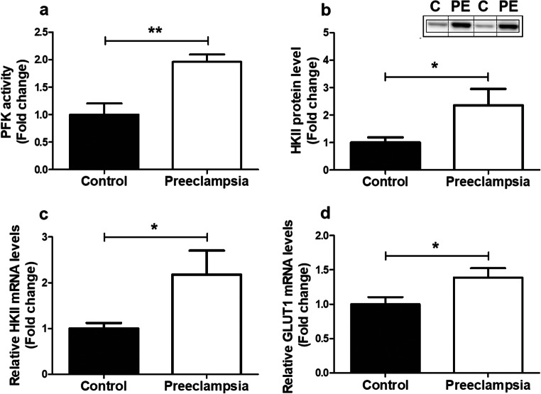
Fig. 3.
Increased expression and activity of key glycolytic enzymes in PE placentae. PFK enzyme activity (a), HKII protein level (b), mRNA expression levels of HKII (c), and Glut-1 (d) were assessed in PE as well as control placentae. Representative immunoblots are shown, and western blots were corrected for total protein loading assessed by Ponceau S staining with adjusted contrast equally applied to the whole photograph. Black boxes around the representative pictures indicate that they were cut from the same western blot. Data is presented as fold change compared to the control placentae and as mean with SEM from n = 11 (controls), n = 12 (preeclampsia) for assessment of mRNA levels and n = 9 (C: controls), n = 8 (PE: preeclampsia) for assessment of the PFK activity and HKII protein levels. Ns: p > 0.05, *p ≤ 0.05, **p ≤ 0.01. PFK, phosphofructokinase; HKII, hexokinase; and GLUT-1, glucose transporter 1

