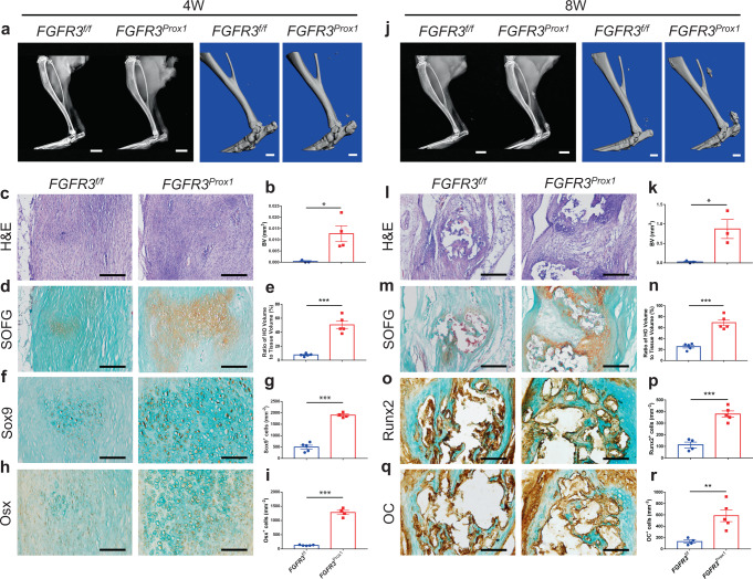
Fig. 3. FGFR3 deletion in LECs aggravates heterotopic bone formation.
a, b Representative X-ray (left) and μCT (right) images of heterotopic bone in the Achilles tendon and quantitative analysis in FGFR3f/f (n = 3) and FGFR3Prox1 mice (n = 4) at 4 weeks after tenotomy. Scale bars, 2 mm for X-ray; 1 mm for μCT. c–e Representative H&E and SOFG images of heterotopic bone and histomorphometry analysis in FGFR3f/f and FGFR3Prox1 mice at 4 weeks after surgery. n = 5 per group. Scale bars, 200 μm. f–i Representative immunohistochemical (IHC) staining of Sox9 (f FGFR3f/f = 5, FGFR3Prox1 = 4) and Osx (h FGFR3f/f = 5, FGFR3Prox1 = 4) and relative quantitative analysis (g, i). Scale bars, 100 μm. j, k Representative X-ray (left) and μCT (right) images of ectopic bone in the tendon and quantitative analysis (k) in FGFR3f/f (n = 3) and FGFR3Prox1 mice (n = 3) at 8 weeks after Achilles tenotomy. Scale bars, 2 mm for X-ray; 1 mm for μCT. l–n Representative H&E and SOFG images of ectopic bone and histomorphometry analysis in FGFR3f/f and FGFR3Prox1 mice at 8 weeks after tenotomy. n = 5 per group. Scale bars, 200 μm. o–r Representative immunohistochemical staining of Runx2 (o FGFR3f/f = 4, FGFR3Prox1 = 5) and OC (q FGFR3f/f = 4, FGFR3Prox1 = 5) and relative quantitative analysis (p, r). Scale bars, 100 μm. All data represent mean ± SEM. *P < 0.05; **P < 0.01; ***P < 0.001 by unpaired two-tailed Student’s t-test.

