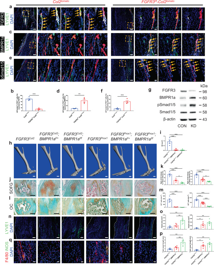
Fig. 5. FGFR3 deficiency in LECs aggravates acquired HO formation via upregulating BMPR1a-pSmad1/5 signaling.
a–f Representative confocal images of repaired Achilles tendon immunostained with FGFR3 (a), BMPR1a (c) or pSmad1/5 (e) (white), LYVE1 (green), and DAPI (blue) in Col2tomato (left) and FGFR3f/f-Col2tomato mice (right) at 8 weeks after surgery and relative quantitative analysis (b, d, f). n = 4 per group. Dashed line boxes indicate tdTomato-labeled LYVE1+ LECs (higher magnification with split channels, right). Scale bars, 100 μm (left); 20 μm (right). g Protein levels of FGFR3, BMPR1a, pSmad1/5, and Smad1/5 in mouse LEC cell line treated with FGFR3-targeted siRNA (KD) or control siRNA (CON). n = 3 per group. h–m Representative μCT (h), SOFG staining (j), and OC immunohistochemistry (l) images of ectopic bone in the Achilles tendon of FGFR3Col2 (n = 4), FGFR3Col2;BMPR1af/+ (n = 5), FGFR3Col2;BMPR1af/f (n = 5), FGFR3Prox1 (n = 4), FGFR3Prox1;BMPR1af/+ (n = 10), and FGFR3Prox1;BMPR1af/f mice (n = 6) at 8 weeks after surgery and relative quantitative analysis (i, k, m). Scale bars, 1 mm for μCT; 200 μm for SOFG and OC IHC. n–q Representative confocal images of Achilles tendon immunostained with LYVE1 (green) and DAPI (blue) (n) in FGFR3Col2 (n = 4), FGFR3Col2;BMPR1af/+ (n = 5), FGFR3Col2;BMPR1af/f (n = 5), FGFR3Prox1 (n = 4), FGFR3Prox1;BMPR1af/+ (n = 5), and FGFR3Prox1;BMPR1af/f mice (n = 6) and relative quantitative analysis (o, p) at 8 weeks after surgery. Scale bars, 100 μm. F4/80 (red), iNOS (green) and DAPI (blue) immunostained Achilles tendon sections (q). Scale bars, 20 μm. White dashed lines indicate outlines of the tendon (a, c, e, n). All data represent mean ± SEM. *P < 0.05; **P < 0.01; ***P < 0.001 by unpaired two-tailed Student’s t-test (b, d, f) or by one-way ANOVA followed by a Tukey’s multiple comparisons test (i, k, m, o, p).

