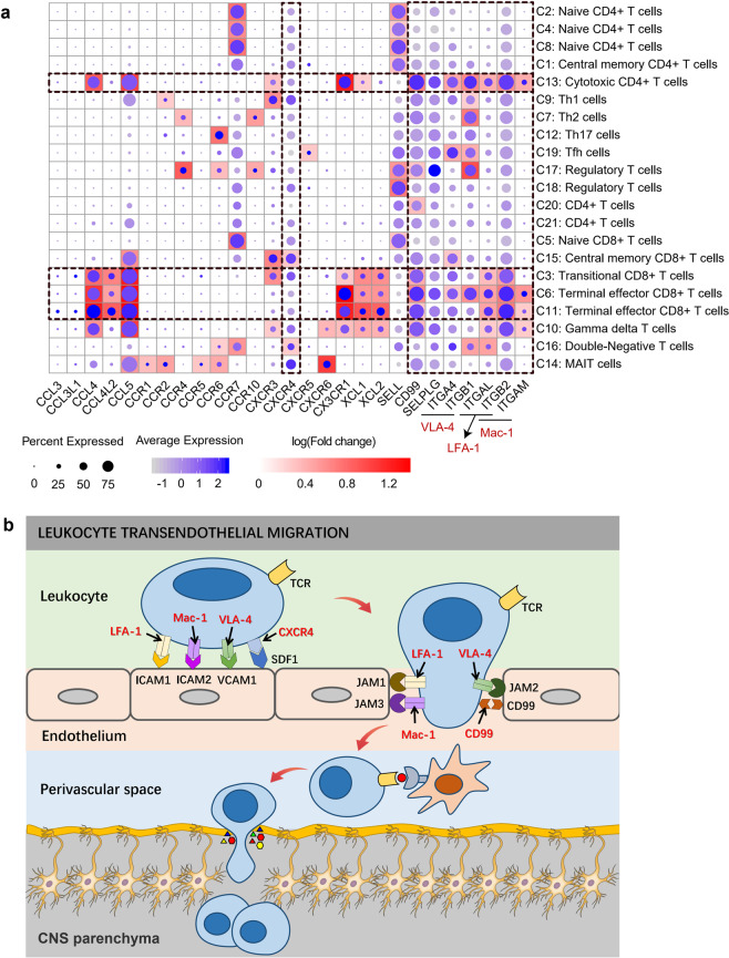
Fig. 6. Cell migration and adhesion-related genes may be involved in lymphocytes penetrating the BBB in the PD.
a A global view of the expression profiling of genes related to cell migration and adhesion in each cluster. The size of the dot corresponds to the percentage of cells expressing the gene in each cluster, and the color represents the average log normalized gene expression. Background heatmap shows the fold change in gene expression between each cluster and other clusters. Fold change was calculated using the function FindAllMarkers in R package Seurat with default parameters. Only positive markers (logFC > 0) in each cluster are shown in the heatmap. b Possible mechanism of cytotoxic T cells passing through the BBB in PD. Depiction of lymphocyte migration across endothelial cells was based on the leukocyte transendothelial migration pathway (KEGG pathway: hsa04670). During inflammation, adhesion molecules, chemokines and their receptors mediate the arrest, polarization, directed crawling and endothelial passage of lymphocytes on endothelial cells. Once T cells cross the BBB endothelium, T cells may recognize their cognate antigens and become reactivated behind the BBB. The amplified inflammatory response leads to parenchymal basement membrane damage. Finally, activated T cells enter the brain parenchyma to participate in central nervous system damage.

