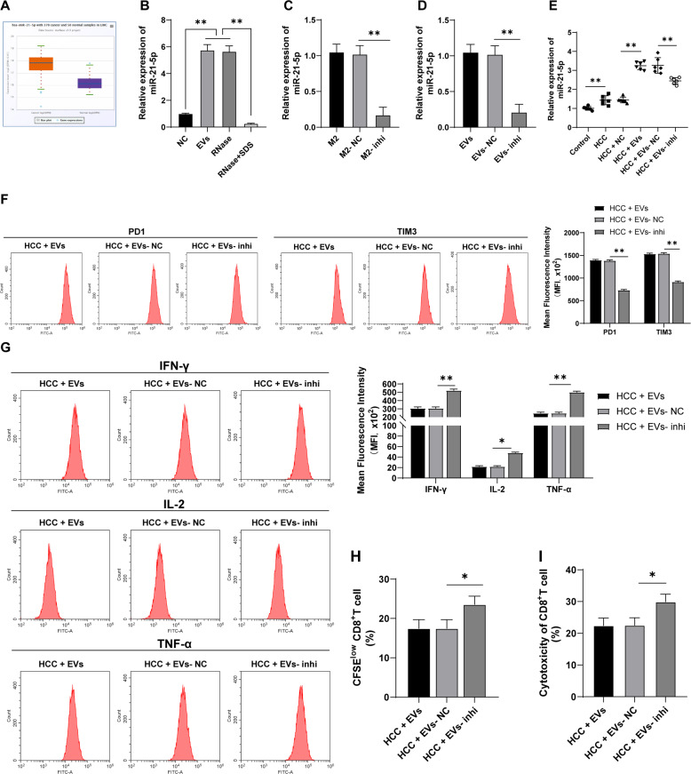
Fig. 2. EVs carried miR-21-5p into the liver tissues of mice.
A miR-21-5p expression in HCC was analyzed through the ECORI Pan-Cer database. B miR-21-5p expression in EVs under different treatments was detected using RT-qPCR. M2 macrophages were transfected with miR-21-5p inhibitor. C, D miR-21-5p expression in M2 macrophages and M2 macrophage-derived EVs was detected using RT-qPCR. E HCC mice were injected with EVs under different treatments and the mice were killed after 20 days to obtain liver tissues, and miR-21-5p expression in mouse liver tissues was detected using RT-qPCR. PBMCs were isolated from the mouse liver and CD8+ T cells were sorted. F The expressions of PD1 and TIM3 on CD8+ T cell surface were detected using flow cytometry. G The expressions of effector cytokines (IFN-γ, IL-2, and TNF-α) were detected using flow cytometry. H The proliferation ability of CD8+ T cells was measured using flow cytometry. I The ability of CD8+ T cells to kill Hep1-6 cells was measured using flow cytometry. N = 6. The cell experiment was repeated 3 times. Data are presented as mean ± standard deviation and analyzed using one-way ANOVA, followed by Tukey’s multiple comparison test, *p < 0.05, **p < 0.01.

