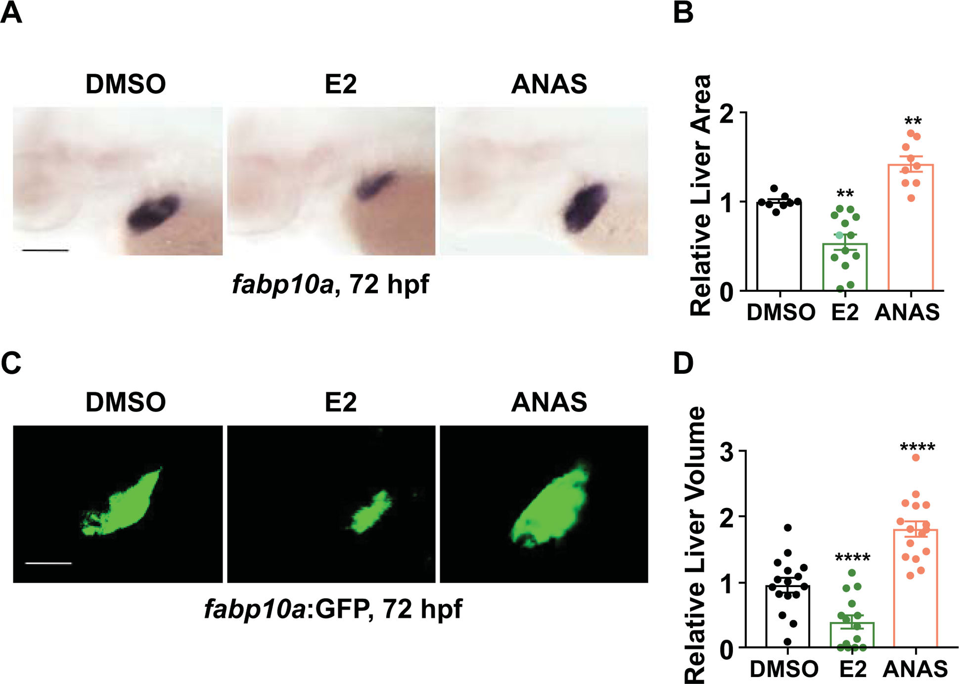
Figure 1. E2 signaling is required for normal embryonic liver formation.

(A) Representative images of zebrafish embryos exposed to DMSO, E2 (10μM), or ANAS (10μM) from 24–72hpf. Liver size assessed by whole mount in situ hybridization (WISH) for liver fatty acid binding protein10a (fabp10a) at 72hpf. (B) Liver marker fabp10a expression area quantified by ImageJ analysis. n ≥ 8, **p<0.01, one-way ANOVA. (C) Representative images of Tg(fabp10a:GFP) embryos exposed to DMSO, E2 (10μM), or ANAS (10μM) from 24–72hpf. (D) Quantification of liver volume in Tg(fabp10a:GFP) embryos exposed to DMSO, E2, or ANAS from 24–72hpf by confocal microscopy analysis at 72 hpf. ****p<0.0001, n ≥ 10, one-way ANOVA. All values represent mean ± SEM, all scale bars, 200μm.
