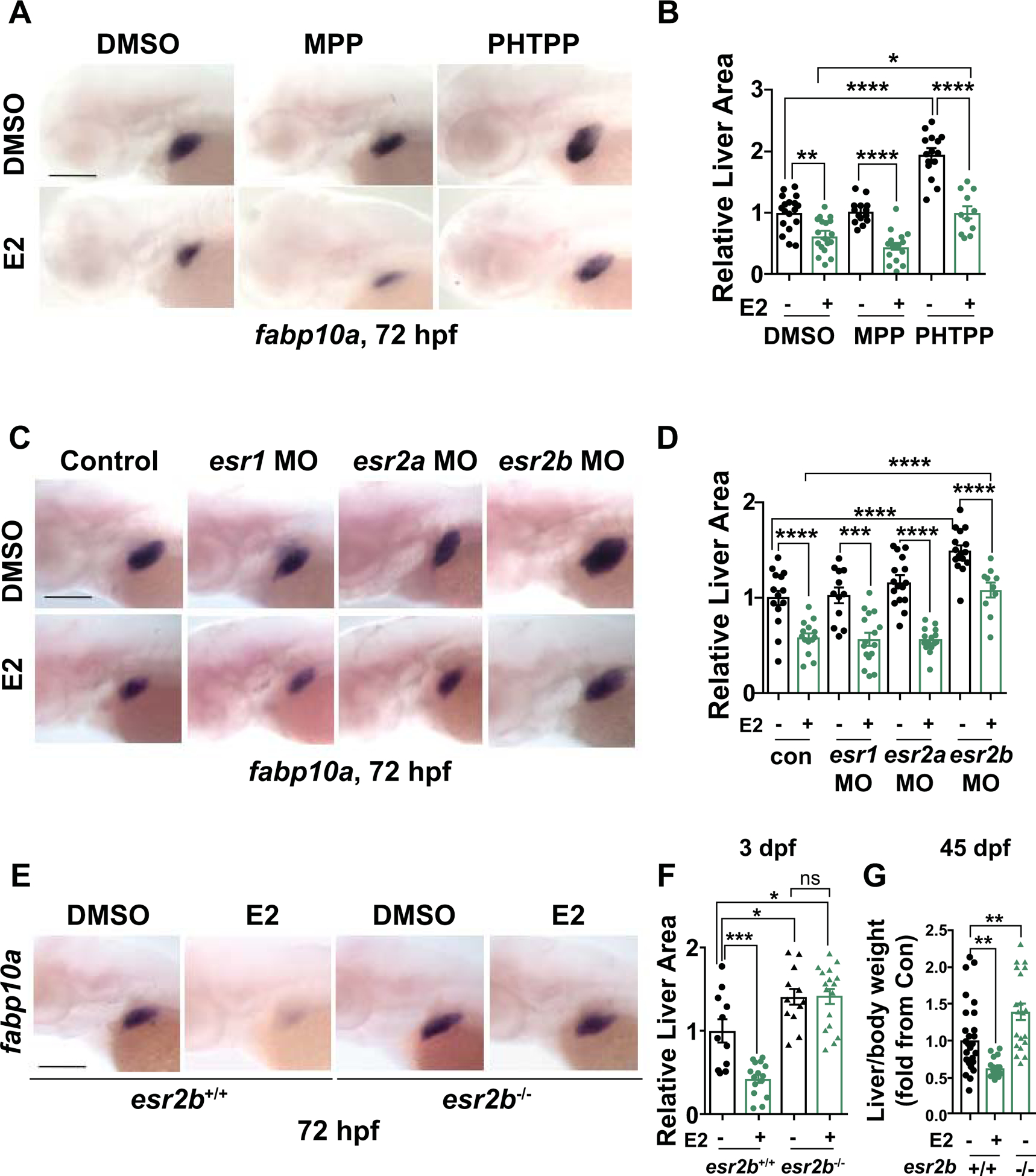
Figure 2. Estrogen receptor 2b mediates impact of E2 on embryonic liver development.

(A) Representative images of WT embryos exposed to DMSO, ESR1 antagonist (MPP), and ESR2 antagonist (PHTPP) alone or together with E2 from 24–72hpf at 72hpf. (B) Quantification of liver size by ImageJ analysis. *p<0.05, **p<0.01, ****p<0.0001, n ≥ 11, one-way ANOVA. (C) Representative images of fabp10a expression at 72hpf in esr1, esr2a, or esr2b morphants exposed to DMSO or E2 from 24–72hpf. (D) Quantification of fabp10a liver area by ImageJ analysis. ***p<0.001, ****p<0.0001, n ≥ 10, one-way ANOVA. (E) Representative images of WISH for fabp10a at 72hpf of esr2b−/− mutants and WT siblings upon exposure to DMSO or E2 from 24–72hpf. (F) Quantification of liver size at 72hpf. ns=not significant, *p<0.05, ***p<0.001, one-way ANOVA. (G) Liver/body weight of 45 dpf esr2b+/+ and esr2b−/− fish exposed to E2 from 24–72hpf, n ≥ 11, **p<0.01, one-way ANOVA. All values represent mean ± SEM, all scale bars = 200 μm.
