Figure 8.
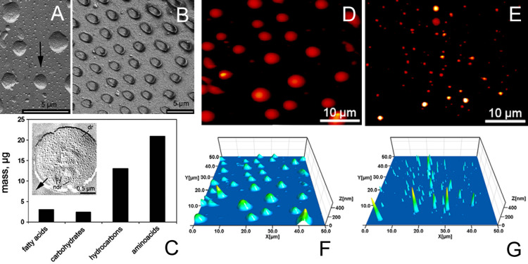
Fluid micro- and nanodrops in animal attachment pads. (A) Carbon–platinum replica of frozen and coated droplets of the fly Calliphora vicina in SEM (black arrow indicates the direction of coating). Please note the pattern of nanodrops on the surface of the major droplets (Figure 8A is from [3] and was adapted by permission from Springer Nature from “Attachment devices of insect cuticle” by S. N. Gorb, Copyright 2001 Springer Nature. This content is not subject to CC BY 4.0). (B) Menisci formed around single terminal contact elements of the setae of C. vicina. The fly leg was frozen in contact with smooth glass, carefully removed, and the fluid residues were viewed in cryo-SEM (Figure 8B is from [241] and was adapted by permission from Springer Nature from “Biological fibrillar adhesives: functional principles and biomimetic applications. In: Handbook of Adhesion Technology” by S. N. Gorb, Copyright 2011 Springer Nature. This content is not subject to CC BY 4.0.). (C) Chemical composition (absolute concentration of substance groups) of the pad secretion of the smooth euplantulae of Locusta migratoria (Figure 8C was adapted from [254], Insect Biochem. Mol. Biol., vol. 32, by W. G. Vötsch; R. Nicholson; Y.–D. Müller; S. Stierhof; S. N. Gorb; U. Schwarz, “Chemical composition of the attachment pad secretion of the locust Locusta migratoria”, pages 1605–1613, Copyright (2002), with permission from Elsevier. This content is not subject to CC BY 4.0). (D–G) Atomic force microscopy (AFM) height images of the footprint droplets of the beetle Coccinella septempunctata (D,F) and the fly Calliphora vicina (E,G). (D) and (E) share the same colour scale. Brighter pixels correspond to higher z values. (F,G) Three-dimensional impressions of the images shown in D and E, respectively (Figure 8D–G was adapted with permission from [262], © 2012 The Company of Biologists Ltd. This content is not subject to CC BY 4.0).

