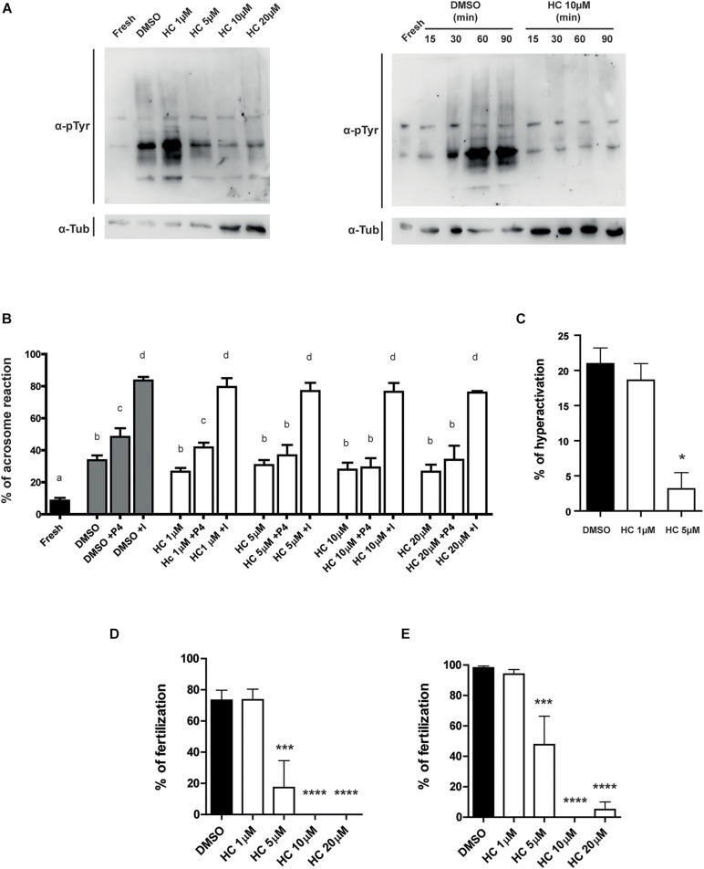
FIGURE 2.
Effect of HC on capacitation-associated functional parameters and in vitro fertilizing ability of sperm exposed to HC during capacitation. (A) Protein tyrosine phosphorylation analysis for protein extracts from non-capacitated sperm (Fresh) or sperm incubated under capacitating conditions in medium containing either different concentrations of HC (1, 5, 10, and 20 μM) or DMSO (vehicle) as control (left panel). Protein tyrosine phosphorylation as a function of incubation time for non-capacitated sperm (fresh) or sperm incubated under capacitating conditions in a medium containing 10 μM HC (right panel). Samples were analyzed by Western blot using a mouse antiphosphotyrosine monoclonal antibody (α-pTyr). In all cases, a representative Western blot from at least three different experiments is shown. (B) Percentage of acrosome reaction determined by Coomassie Brilliant Blue staining in cauda epididymal sperm incubated under capacitating conditions for 90 min in medium containing different concentrations of HC (1, 5, 10, and 20 μM) and exposed to either progesterone (P4) or Ca2+ ionophore (I) during the last 15 min of incubation. Non-capacitated (fresh) sperm were used as control. Means not sharing the same letters are significantly different. (C) Percentage of hyperactivation evaluated by CASA for sperm incubated under capacitating conditions for 90 min in a medium containing 1 or 5 μM HC or DMSO (vehicle) as control. (D,E) Percentage of fertilization obtained using cauda epididymal sperm incubated for 90 min in capacitating medium containing different concentrations of HC (1, 5, 10, and 20 μM) or DMSO (control) and then coincubated with either Cumulus Oocytes Complexes (COC) for 3 h (D) or ZP-free oocytes for 1 h (E). At the end of incubations, fertilization was evaluated. Eggs were considered fertilized when at least one decondensing sperm nucleus or two pronuclei were observed in the egg cytoplasm and the percentage of fertilized eggs was determined. Data are mean ± SEM of at least n = 3 independent experiments; *p < 0.05; ***p < 0.0005, and ****p < 0.0001 vs. DMSO.

