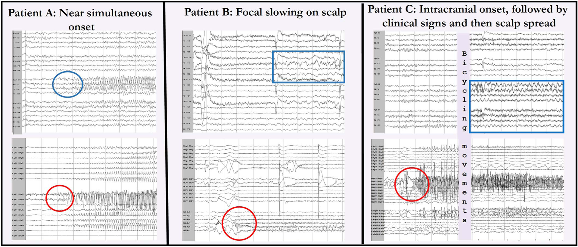
FIG. 1.

Comparison of select simultaneous scalp and intracranial EEGs. In patient A, the intracranial onset (bottom circle) in the right hippocampus was seen near simultaneously on scalp (top circle) in the right temporal chain. For patient B, an intracranial right lateral temporal onset (circle) was followed by 5 seconds of polymorphic right temporal slowing (box), which was not sustained and not different from the interictal slowing. In patient C, there was an intracranial right hippocampal onset (circle) with no scalp EEG finding followed by clinical signs and then rhythmic diffuse right hemispheric theta slowing seen on scalp (box).
