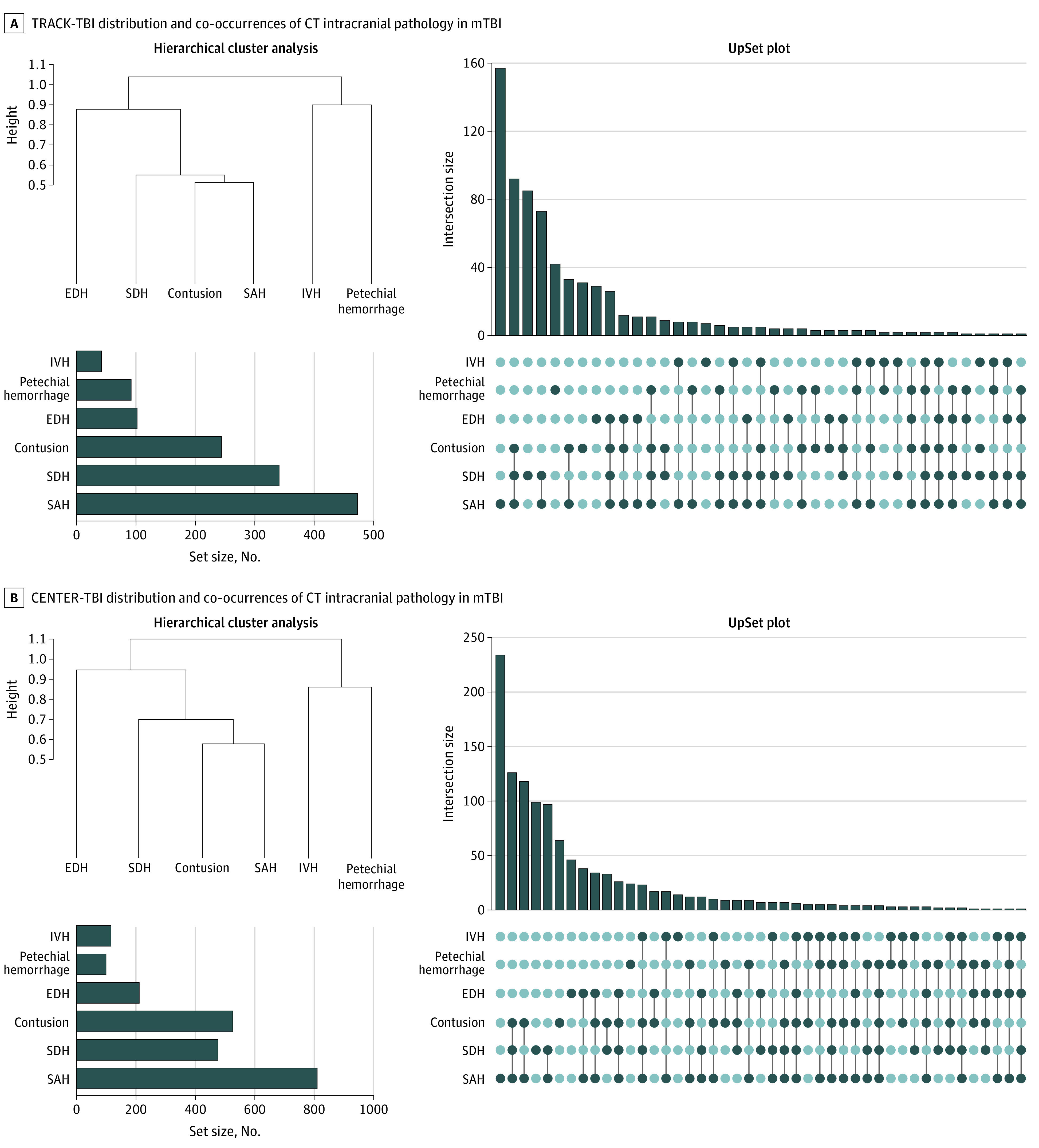
Figure 2. Distribution and Co-occurrences of Intracranial Pathology on Computed Tomography (CT) in Mild Traumatic Brain Injury (mTBI) by Cohort.

A, Distribution of National Institute of Neurological Disorders and Stroke TBI Neuroimaging Common Data Elements (CDEs) in participants 17 years and older with Glasgow Coma Scale scores of 13 to 15 (n = 1935) in the Transforming Research and Clinical Knowledge in TBI (TRACK-TBI) study. An UpSet plot shows that the most common pattern of acute intracranial hemorrhage is isolated subarachnoid hemorrhage (SAH), which constitutes 157 of 715 (22.0%) of all CT examinations showing intracranial hemorrhage. (Hierarchical cluster analysis demonstrates clusters of CT abnormalities. A dendrogram shows the distance at which the cluster was formed along the vertical axis, with 3 clusters: contusion, SAH, and/or subdural hematoma (SDH); intraventricular hemorrhage (IVH) and/or petechial hemorrhage; and epidural hemorrhage (EDH). The bar graph in the lower left corner shows that the most common acute intracranial abnormality was SAH (in 473 of 1935 patients [24.4%]), followed by SDH (341 [17.6%]), brain contusion (244 [12.6%]), EDH (102 [5.3%]), petechial hemorrhage (92 [4.8%]), and IVH (42 [2.2%]). B, Distribution of CDEs in participants 17 years and older with Glasgow Coma Scale scores of 13 to 15 (n = 2594) in the Collaborative European NeuroTrauma Effectiveness Research in Traumatic Brain Injury (CENTER-TBI) study. An UpSet plot shows that the most common pattern of acute intracranial hemorrhage is isolated SAH, which constitutes 234 of 1175 (19.9%) of all CT examinations positive for intracranial hemorrhage. Hierarchical cluster analysis shows clusters of CT abnormalities. A dendrogram shows the distance at which the cluster was formed along the vertical axis. The most common acute CT finding was SAH (810 of 2594 patients [31.2%]), followed by brain contusion (526 [20.3%]), SDH (476 [18.4%]), EDH (211 [8.1%], IVH (116 [4.5%]), and petechial hemorrhage (99 [3.8%]).
