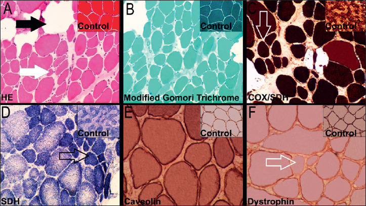
Figure 1.

Quadriceps muscle biopsy with features of dystrophy. A) Muscle fibres showing variation in size and atrophic fibres (white arrow) surrounded by endomysial connective tissue/adipose tissue (dark arrow) (Hematoxylin and Eosin, 10x); B) Muscle section with variation in fibre size, internal nuclei, increased endomysial connective tissue and adipose tissue (modified Gomori trichrome, 10x); C) and D) Oxidative enzymes showing variation in fibre size with slight predominance of type 1 fibres (open arrow) [cytochrome c oxidase/succinate dehydrogenase (COX/SDH) and succinate dehydrogenase (SDH), respectively; 10x]. (E) Immunolabelling of caveolin-3 using peroxidase label in the same muscle (20x). (F) Immunolabelling of dystrophin revealing some fibres with weak and uneven (open arrow; 20x) labelling compared with controls.
