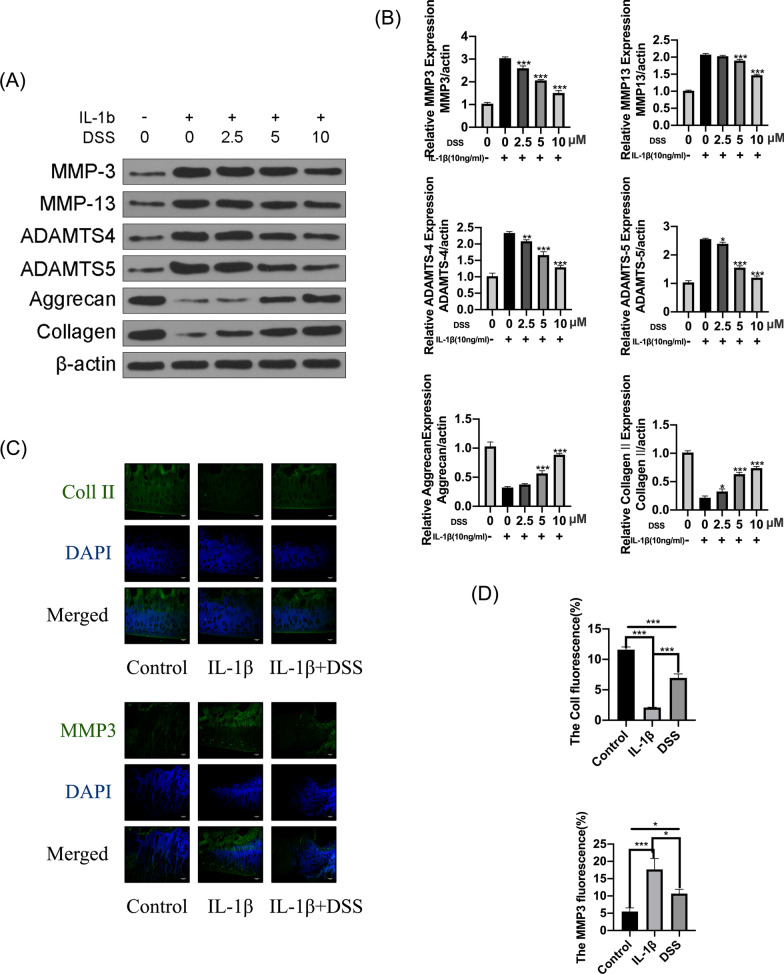
Fig. 3.
DSS protected chondrocytes against IL-1β-induced ECM degradation. The chondrocytes were treated with 2.5, 5, and 10 μM DSS for 24 h and then stimulated by 10 ng/ml IL-1β for 2 h. A, B Protein levels of MMP3, MMP13, ADAMTS4, ADAMTS5, aggrecan, and collagen II in chondrocytes (A), and their quantification by the Quantity ONE software (B). C, D Representative fluorescence images of collagen II and MMP3 detected by immunofluorescent staining assay (C) and the percentage of cell immunofluorescence in chondrocytes that were pretreated with 10 μM DSS for 24 h and stimulated by 10 ng/ml IL-1β for 2 h (D). Scale bar = 50 μm. *p < 0.05, **p < 0.01, and ***p < 0.001

