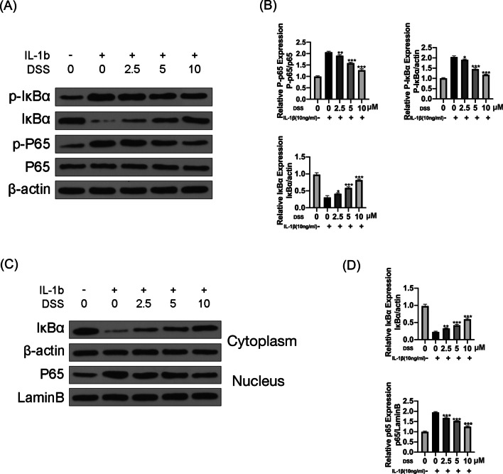
Fig. 4.
DSS inhibited IL-1β-induced activation of the NF-κB pathway in chondrocytes. The chondrocytes were treated 2.5, 5 and 10 μM DSS for 24 h and then induced with or without 10 ng/ml IL-1β for 2 h. The protein levels of p65, p-p65, IκBα, and p-IκBα were detected by Western blot (A) and quantitative analysis (B). The protein levels of IκBα in the cytoplasm and p65 in the nucleus of chondrocytes were detected by Western blot (C) and quantitative analysis (D). *p < 0.05, **p < 0.01, and ***p < 0.001

