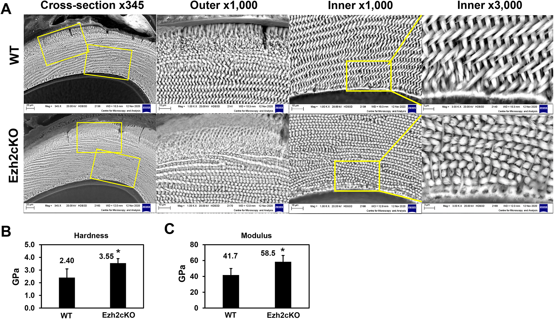
Fig. 2.

(A) The cross-section images at the eruption site of incisors from WT or Ezh2cKO mice, obtained by scanning electron microscopy. Yellow boxes indicate the outer or inner enamel areas shown magnified with indicated powers. The average values of (B) hardness and (C) Young’s modulus properties of the enamel from four WT and six cKO mandibular incisor tips were obtained using nanoindentation tests. A minimum of seven and a maximum of twenty indentation tests per sample were carried out on the enamel area. Error bars indicate standard deviations. *p < 0.05.
