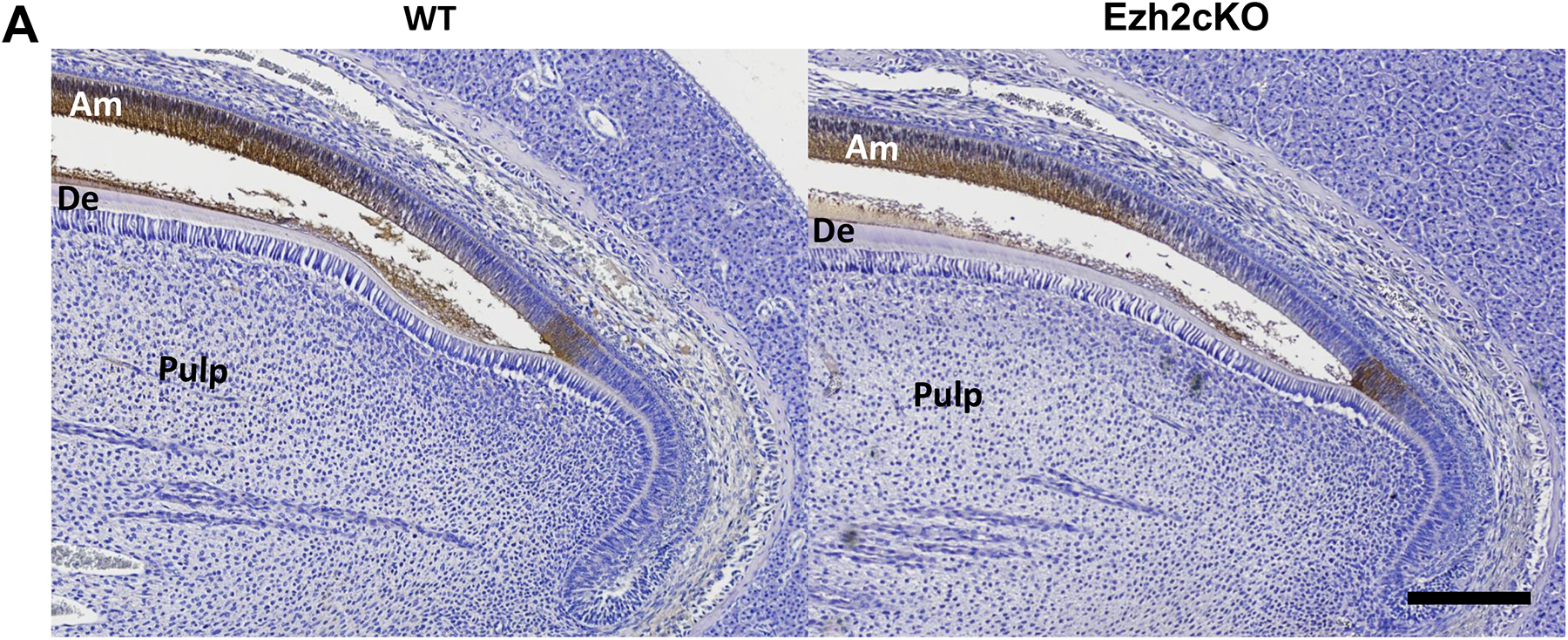
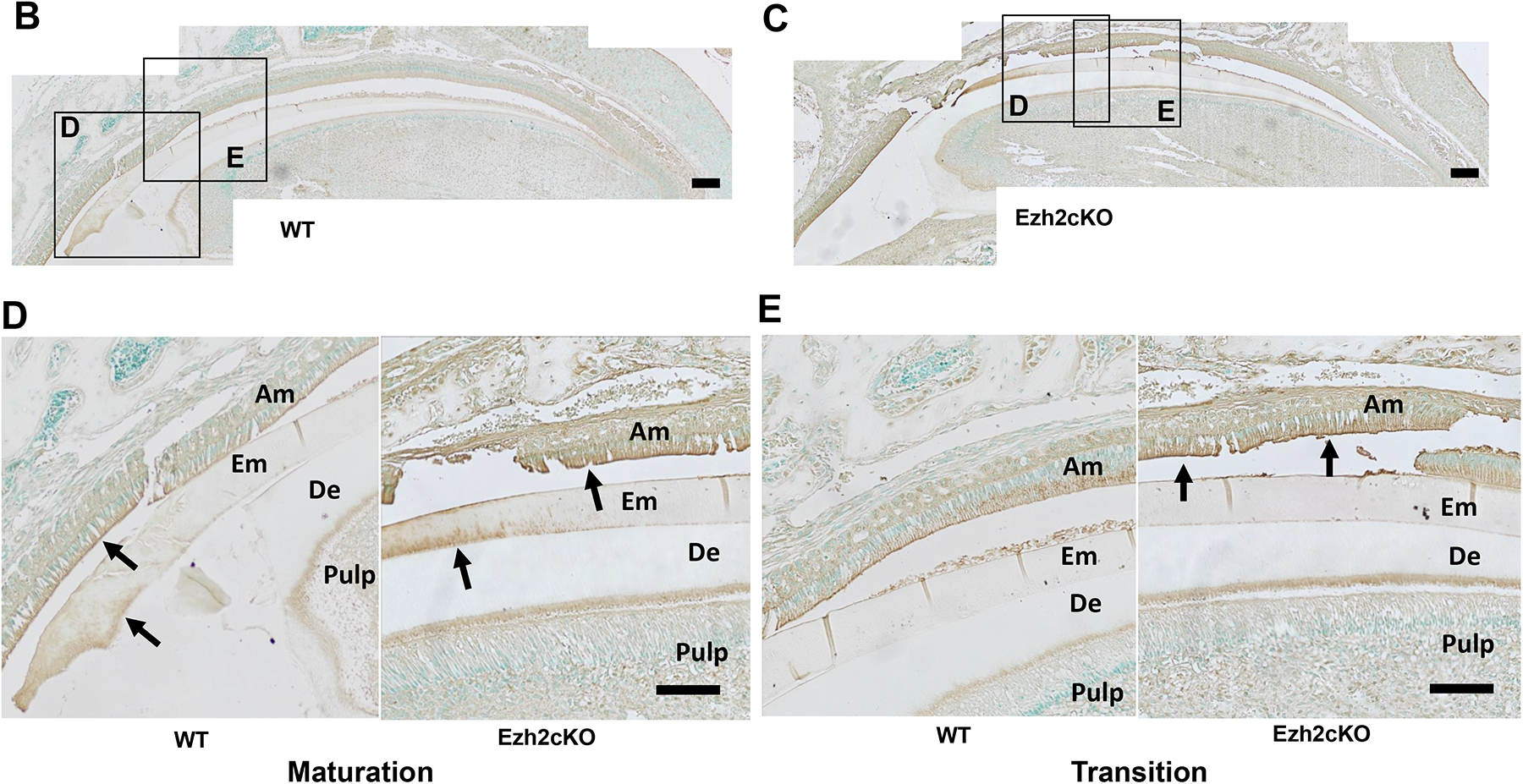
Fig. 4.


(A) Ameloblastin expression in the maxillary incisor from WT (left) or Ezh2cKO mouse (right). Sagittal sections of the incisor were stained with hematoxylin and DAB using HRP-conjugated anti-ameloblastin antibody (brown). Portions close to the cervical loop are shown. Ameloblastin signal is shown to be localized in ameloblasts and the enamel matrix surface. Scale bar = 100 μm. (B, C) KLK4 expression in maxillary incisor from (B) WT or (C) Ezh2cKO mouse. Sagittal sections of the incisor were stained with methyl green and DAB using HRP-conjugated anti-KLK4 antibody (brown). Scale bar = 200 μm. Magnified images (D) and (E) show maturation stage and transition stage ameloblasts from WT and knockout mouse, respectively. Arrows indicate residual KLK4 protein stained with DAB. Am: ameloblast, De: dentin, EM: enamel matrix. Scale bar = 100 μm.
