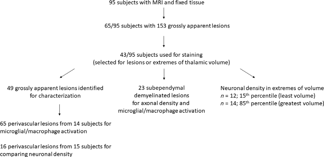
FIGURE 1:
Flowchart of the number of subjects and tissue specimens used for the study. Subsets of tissue sections were used to stain for demyelinating lesions and characterize lesions based on microglial/macrophage activity and neuronal/axonal density. Subjects were also selected for extremes of thalamic volume identified on magnetic resonance imaging (MRI; 15th and 85th percentiles).

