FIGURE 2:
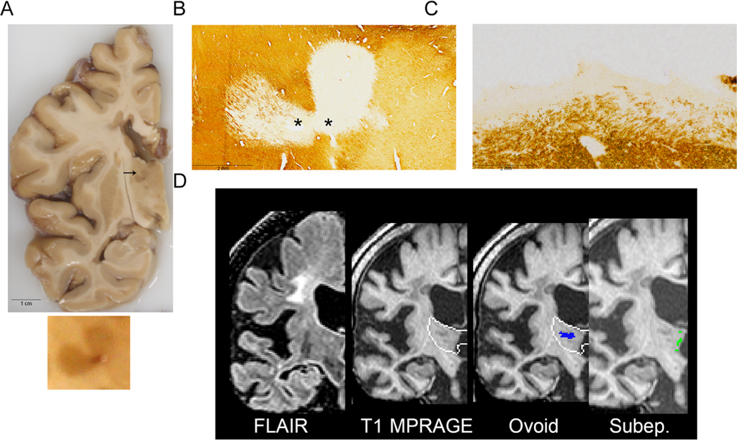
Representative case of a subject with grossly apparent hyperpigmented thalamic lesions on a 1 cm fixed coronal slice (A); enlarged lesion shown below. Both ovoid (B) and subependymal (C) lesions are noted using PLP immunohistochemistry for myelin; asterisk (*) denotes location of vessel. Magnetic resonance imaging (MRI) coronal images, with thalamus depicted with a white outline, (D) depict fluid-attenuated inversion recovery (FLAIR) and T1-Magnetization Prepared Rapid Acquisition of Gradient Echo (MPRAGE) sequences as well as ovoid (blue) and subependymal (Subep.; green) manually segmented lesions. The heart-shaped hyperpigmented lesion straddling the dorsomedial and ventrolateral nuclei is indicated with an arrow and an enlarged inset of the lesion is below (A). The lesion is demyelinated (B) and labeled as ovoid/perivascular (“Ovoid”) on MRI T1 MPRAGE (D). Subependymal lesions (C) were not identified on gross examination (A) and subtle on MRI (D; labeled “Subep.”). PV, perivascular; PLP, proteolipid protein.

