
Figure 3. Pediatric TBI does not result in an overt lesion or loss of NeuN immunoreactivity within the mPFC.
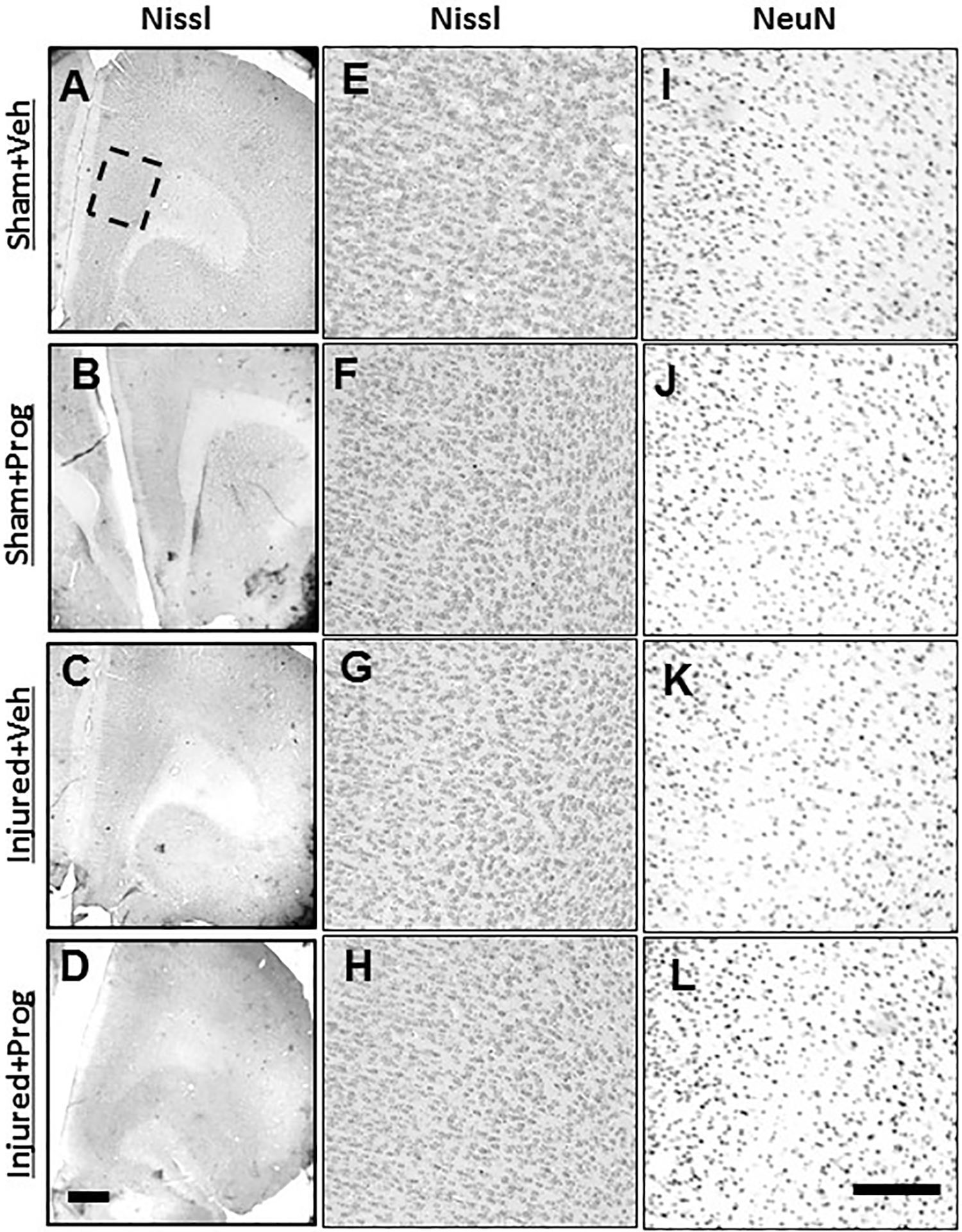
Representative images illustrating Nissl staining in coronal sections containing the mPFC (A-H) and NeuN immunoreactivity (I-L) from sham- (A,B,E,F,I,J) and brain-injured rats (C,D,G,H,K,L) that received sesame oil (Veh; A,C,E,G,I,K) or progesterone (Prog; B,D,F,H,J,L), at 4 weeks after sham- or brain-injury. Boxed area in panel A represents the prelimbic region of medial PFC depicted at higher magnification in panels E-L. Scale bar in panel D = 200 μm for panels A-D and in panel L = 50 μm for panels E-L.