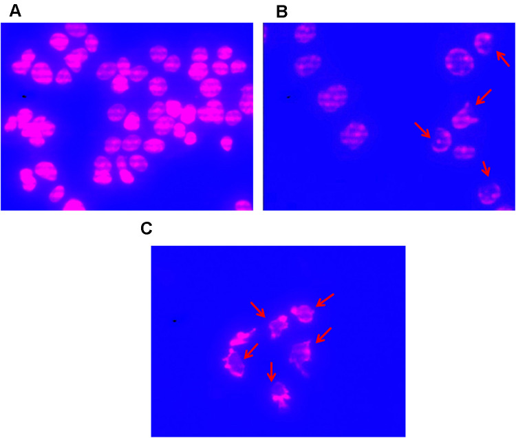
Figure 3.
Changes of MCF7 cells form figure. (A) MCF7 cells were exposed extracts of A. muricata L. for 0 h; (B) MCF7 cells after treated with A. muricata L. Extract for 6 h; (C) MCF7 cells after treated with A. muricata L Ethyl acetate fraction for 6 h. MCF7 cells observed under fluorinated ZEISS Apotome.2 (Carl Zeiss, Germany) microscopes with Cy3 and DAPI filters. MCF7 cells form after treatment with A. muricata L. either extracts or ethyl acetate fractions to be inconsistent and rupture to parts (indicated by a red arrow).

