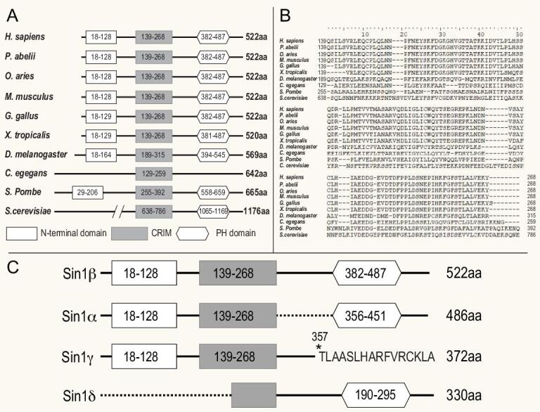
Figure 2.

Sequence alignment of evolutionarily conserved Sin1 proteins. (A) A diagram showing the evolutionarily conserved domains in Sin1 proteins among different species. Empty rectangle, N-terminal region; filled rectangle, conserved region in the middle (CRIM); hexagon, PH domain. (B) Sequence alignment of the CRIM domain of Sin1 from different species. (C) Alternative splicing isoforms of human Sin1.
