Figure 8.
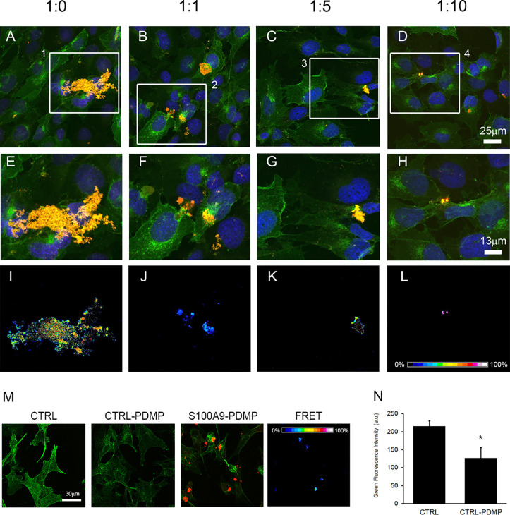
Immunolocalization of S100A9 aggregates on the SH-SY5Y plasma membrane. Confocal microscopy imaging of SH-SY5Y cells exposed for 24 h to 20 μM S100A9 aggregated for 48 h in the absence or in the presence of different molar ratios of S100A9 to OleA as indicated in the figures. The cell membranes were stained with Alexa 488-conjugated CTX-B (green fluorescence); cell nuclei were stained with Hoechst 33342 (blue fluorescence), and protein aggregates were stained with anti-S100A9 antibodies followed by treatment with Alexa 568-conjugated antirabbit secondary antibodies (red fluorescence). The mergence of the channels is shown in (A–D). Zoomed areas from (A–D) are shown in (E–H). FRET efficiency in those areas is shown in (I–L). (M) S100A9 do not bind to membrane rafts in the SH-SY5Y cells pretreated with an inhibitor of the GM1 synthesis as revealed by the lack of FRET efficiency. The control corresponds to the untreated cells. The SH-SY5Y cells were pretreated with 10 μM PDMD for 48 h (indicated as CTRL-PDMP) to reduce GM1 on the cell surface and then incubated for 24 h with 20 μM S100A9 fibrils (in monomer concentration). (N) Quantification of green fluorescence per cell, indicating the reduction of GM1 in cell membranes. Error bars indicate the standard error of three independent experiments. *p < 0.001 versus control.

