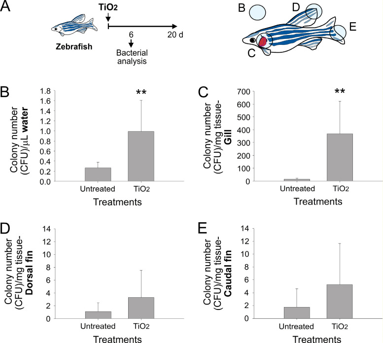
Fig 3. Analysis of the relative number of bacteria found in the water and zebrafish tissue samples.
(A) Experiment outline and (B) sampling positions for bacterial culture. The culturable bacterial number in (B) the water, (C) zebrafish gill, (D) dorsal fin, and (E) caudal fin after analysis with the plating method. ** P < 0.01, compared with the untreated control groups.

