
Figure 1.
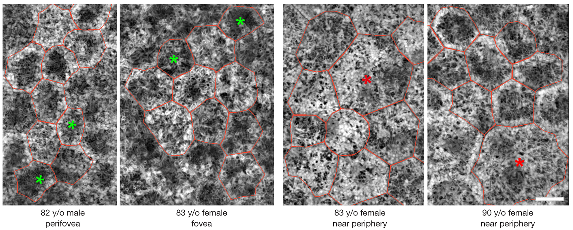
Retinal pigment epithelium (RPE) cells with low/high granule load. The colored asterisks mark the cells with low (green) and high (red) amounts of granules. Individual RPE cells are red-rimmed. Structured Illumination Microscopy (summation of the whole image stack). Scale bar: 10 μm.