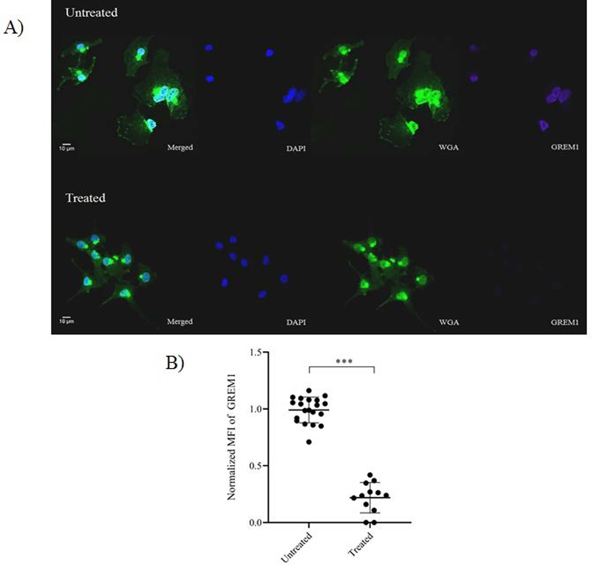
Figure 4. Confocal microscopy of GREM1 in cumulus cells isolated from women with normal BMI following treatment with obese follicular fluid.

A) Representative image depicting GREM1 intensity in cumulus cells following treatment with obese follicular fluid. Nucleus was stained with DAPI (blue). Glycocalyx on the plasma membrane was stained with Alexa Fluor-488 conjugated wheat germ agglutinin (green). GREM1 was stained with Alexa Fluor-647 (purple). The bar indicates 10 μm. B) Comparison of normalized MFI of GREM1 between untreated and obese follicular fluid treated cumulus cells. Data were compared by paired t test (N=3 women with 9–12 images per treatment condition). *** indicates p<0.001.
