
Fig 5.
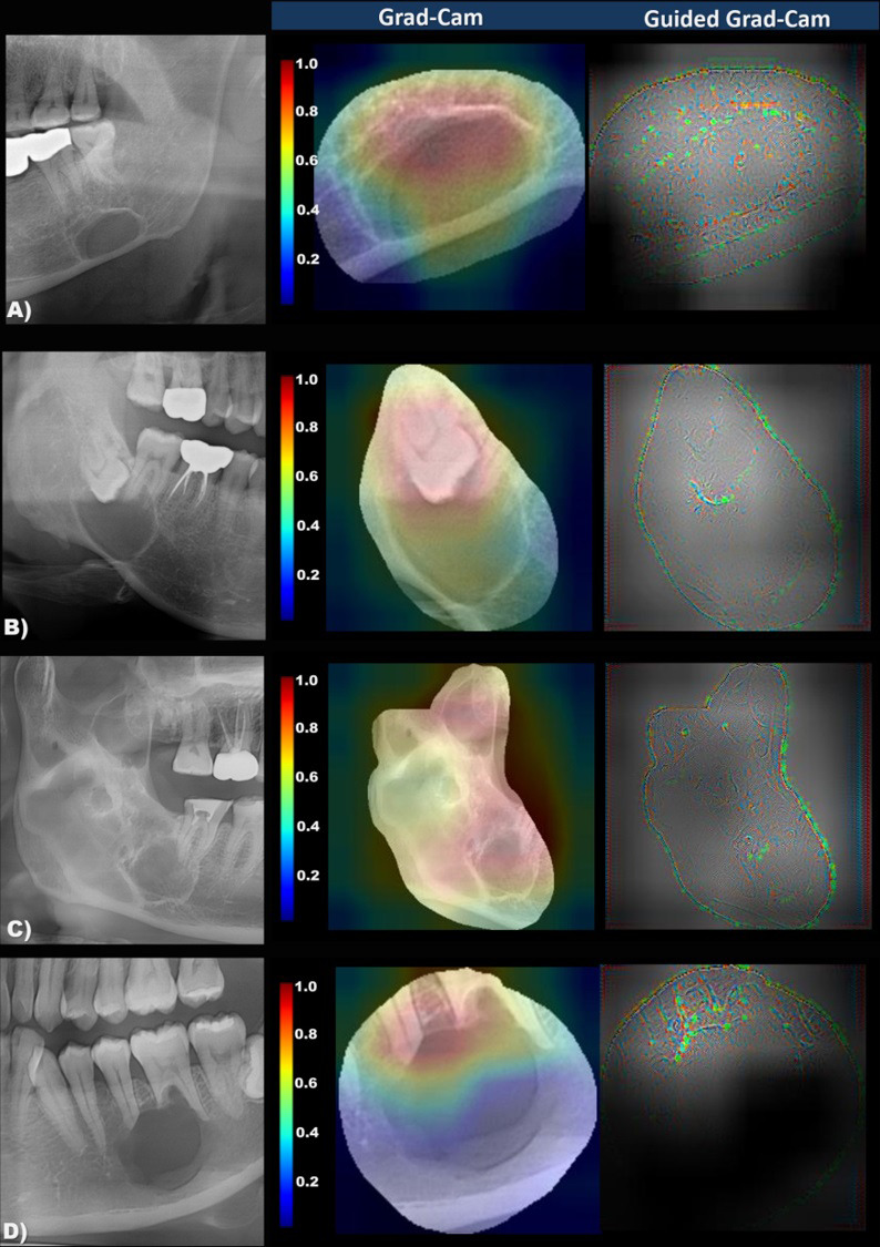
The panoramic image and the importance-weighted visualization image of classification criteria (Grad-Cam and Guided Grad-Cam) in Stafne’s bone cavity (A), dentigerous cyst (B), odontogenic keratocyst, (C), and ameloblastoma (D). Note that the important degree of imaging features for Stafne’s bone cavity classification is color-coded from red (highly-weighted) to blue (less-weighted). The model visualizes the empty internal area and mandibular inferior cortex in Stafne’s bone cavity, while tooth-bearing, multiple locules, and root resorption are well recognized in cysts and tumors.