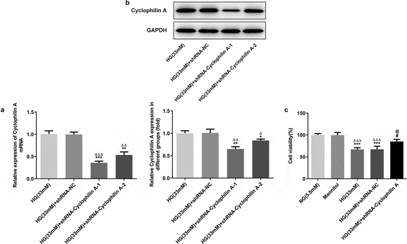
Figure 2.

The effects of cyclophilin A silencing on the cell viability in HG-treated pancreatic β-cells.
The cyclophilin A level evaluated by PCR (a) and Western blot (b) in the study groups. The cell viability level in the study groups (c).*P < 0.05 and **P < 0.01 and ***P < 0.01 vs. HG (33 mM) group, ΔP < 0.05 and ΔΔP < 0.01 and ΔΔΔP < 0.01 vs. HG (33 mM) +shRNA-NC group (A and B); ***P < 0.001 vs. NG group, ΔΔΔP < 0.001 vs. Mannitol group, #P < 0.05 vs. HG (33 mM) group, @P < 0.05 vs. HG (33 mM) + shRNA-NC group (C)
