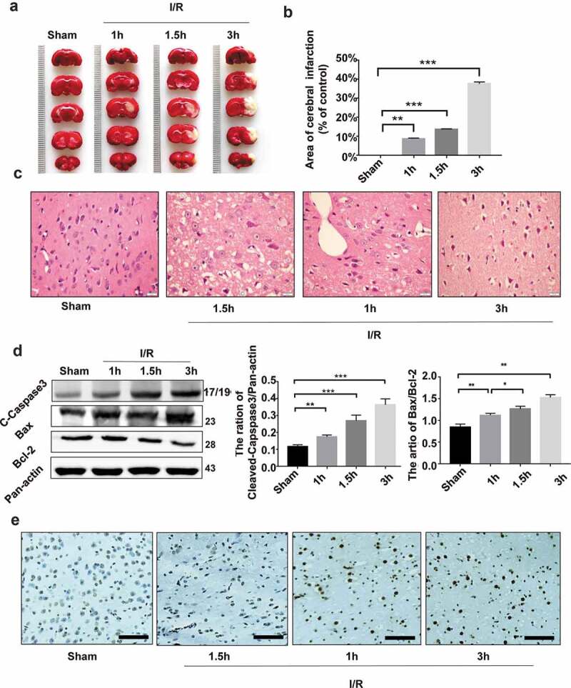
Figure 1.

Ischemia/reperfusion injury causes infarction and apoptosis of rat brain tissues. The rat middle cerebral artery occlusion (MCAO) model was established as detailed in Methods. The MCA was occluded for 1, 1.5 and 3 h, respectively, followed by reperfusion for 24 h. (a) The photographs of the brain samples of rats undergoing sham operation or MCAO. (b) The infarct ratio of 2% TTC stain in each group. Data are presented as mean ± SD, n = 3. **P < 0.01 and ***P < 0.001 vs. the sham group. (c) H&E staining of the infarct marginal zone in the cerebral cortex. Bar = 20 μm. (d) Immunoblotting assays of cleaved caspase 3 and Bac/Bcl-2 in rat brain tissues undergoing ischemia/reperfusion injury. A representative immunoblot is shown (left) and Data are presented as mean ± SD, n = 3. *P < 0.05, **P < 0.01 and ***P < 0.001 vs. the sham group. (e) TUNEL staining of the infarct marginal zone in the brain cortex. Bar = 10 μm.
