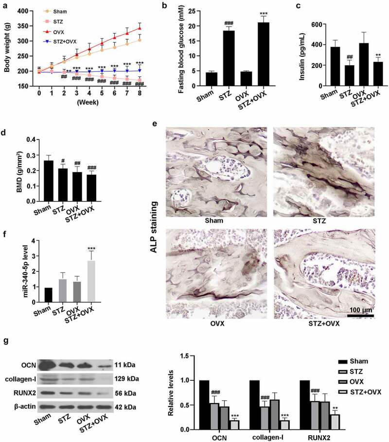
Figure 1.

The rat model of diabetic osteoporosis was established. (a-c) Diabetes was mediated by STZ in rats. Subsequently, bilateral OVX was carried out. The rats were fed for 8 weeks, and body weight was detected once a week. Then the animals were fasted overnight, and fasting blood glucose and blood insulin contents were assessed using the commercial kits. Finally, all the animals were sacrificed, and rat femur tissues were collected for following experiments. (d) Detection of BMD. (e) ALP staining was performed in femur tissues. Scale bar = 100 μm. (f) Measurement of miR-340-5p expression by qRT-PCR. (g) Evaluation of OCN, collagen-I, and RUNX2 levels with immunoblotting. β-actin was used as the internal reference. STZ, streptozotocin; OVX, ovariectomy; BMD, bone mineral density; ALP, alkaline phosphatase; OCN, osteocalcin. Data were expressed as means ± SD (N = 6 per group). #P < 0.05, ##P < 0.01, and ###P < 0.001 versus sham group; **P < 0.01 and ***P < 0.001 versus OVX group
