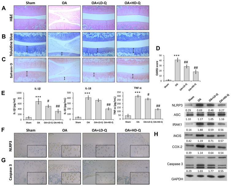
Figure 1.
Quercetin alleviated OA progression and suppressed inflammation in vivo. Rats were assigned to 4 separate groups: a sham, OA, OA + LD-Q, and OA + HD-Q group. (A–C) The degree of cartilage destruction and joint spacing in each group was evaluated at 12 weeks by H&E staining (A), toluidine blue (B) and safranin O staining (C). Arrow in (B and C) showed the distribution of chondrocyte. (D) Osteoarthritis Research Society International (OARIS) scores for articular cartilage in the 4 groups. (E) The levels of pro-inflammatory cytokines (IL-1β, IL-18, and TNF-α) were measured by ELISA. (F and G) Immunohistochemical staining for NLRP3 expression (F) and Caspase 3 (G). (H) The levels of NLRP3, ASC, IRAK1, iNOS, COX-2 and caspase 3 proteins were measured by Western blotting. Data represent mean ± standard deviation. ***p < 0.001, compared with sham; #p < 0.05, ##p < 0.01, compared with OA.

