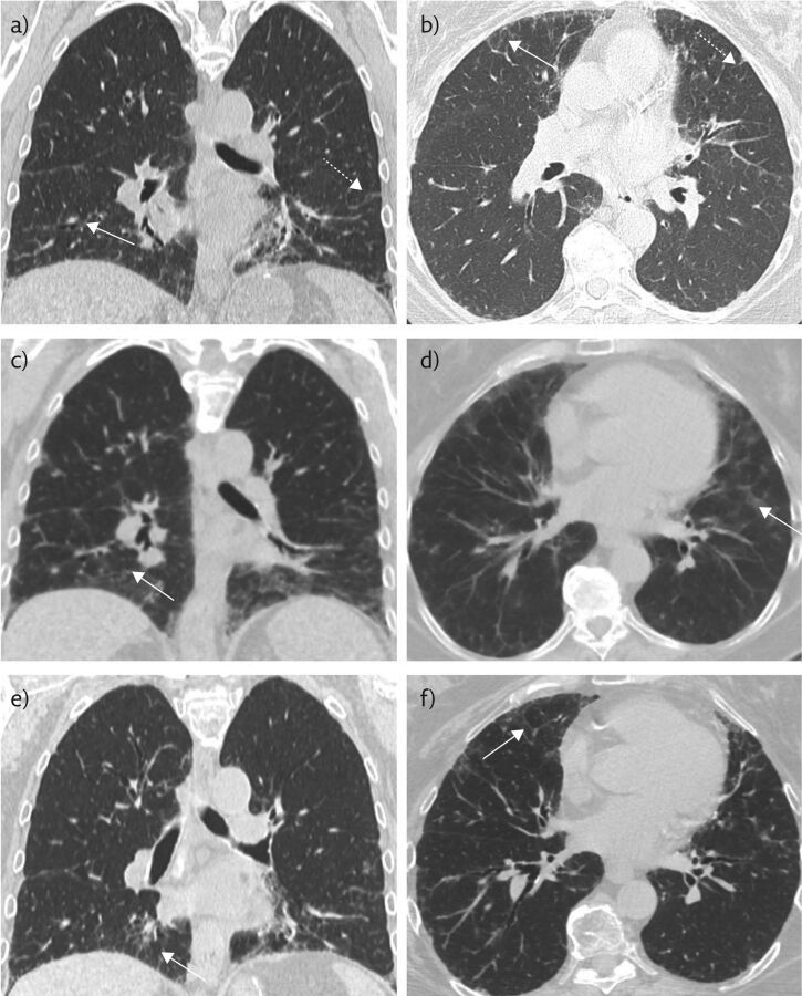
Figure 1.
a, b) Non-contrast chest CT 8 months prior to presentation, when the patient was mildly symptomatic, in the coronal and axial planes, respectively. These show mild basilar-predominant peripheral reticulations (dashed white arrows) and minimal GGOs (solid white arrows). c, d) Representative coronal and axial images during acute exacerbation, the solid white arrows indicate GGOs. e, f) Representative coronal and axial images after discontinuation of nitrofurantoin and initiation of steroid therapy, with near-complete resolution of GGOs and peripheral reticulations.

