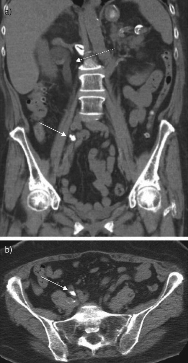
Figure 2.

Representative a) coronal and b) axial images from a non-contrast chest CT of the abdomen and pelvis of this 64-year-old female obtained to evaluate for renal calculus. An obstructive calculus is seen in the distal right ureter (solid white arrow) with proximal hydroureteronephrosis (dotted white arrow).
