
Fig. 12.
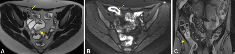
( A ) MRE T2 axial HASTE sequence image in a patient with Crohn's ileitis revealing ileal stricture (thin arrow) and upstream small bowel dilation (large arrowhead). ( B ) MRE trace diffusion-weighted sequence image in the same patient revealing high-signal intensity in the ileus (arrow) consistent with active Crohn's disease. ( C ) MRE T1 coronal VIBE Dixon method sequence image post contrast in the same patient with Crohn's ileitis revealing ileal mural hyperenhancement (thick arrow), thickening, and “comb sign” (thin arrow).