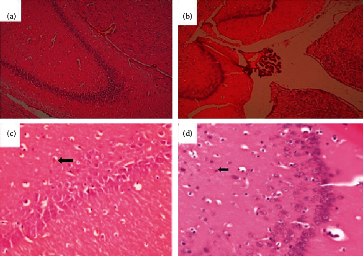
Figure 11.

Brain of a rat that was orally administered 1,000 mg/kg methanolic extract of Psychotria ankasensis (H&E x 100 (a), x 400 (c)). Brain structure is normal. However, while most neurons are normal in structure, a few scattered ones show condensed eosinophilic cytoplasm and dark, shrunken nucleus. This change is consistent with acute ischemic neuronal injury, (C) arrow). The brain of a rat administered with vehicle as control (H&E, x100 (b), x400 (d)) shows that majority of neurons and glial cells are well preserved, and the organ is predominantly normal. One or a few neurons show acute ischemic neuronal injury characterized by the condensed nucleus and reduced cytoplasm that stains more eosinophilic (pinker, image D, arrow).
