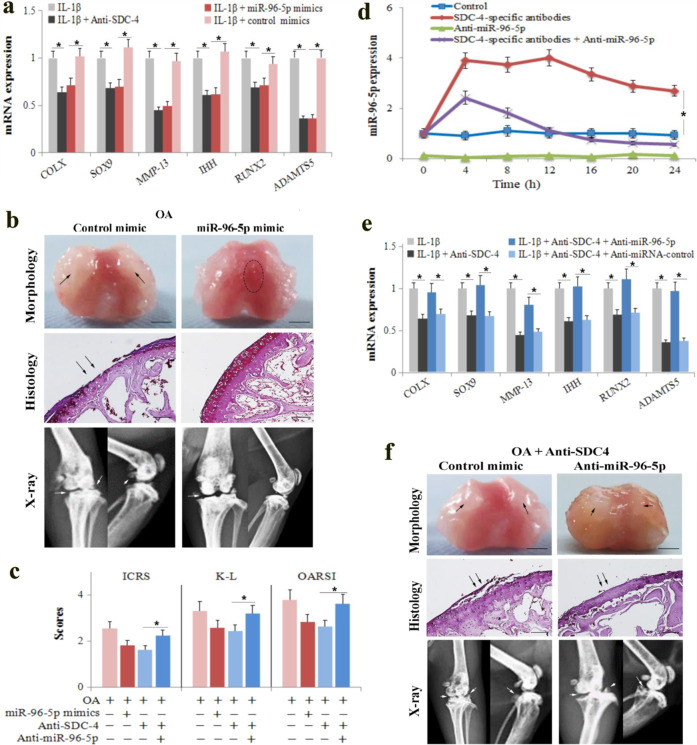
Fig. 3. MiR-96-5p mediates the effects of SDC-4-specific antibodies on murine osteoarthritis and chondrocyte hypertrophy.
a Primary mouse chondrocytes were transfected with miR-96-5p mimics or control mimics for 24 h, pretreated with the syndecan-4-specific antibody for 2 h, and then stimulated with or without recombinant human IL-1β for 4 h. The expression of COL-X, SOX9, MMP-13, IHH, RUNX2, and ADAMTS5 was determined by RT-PCR analyses in chondrocytes. b OA mice were administered 10 μl miR-96-5p mimics or miRNA scrambled controls in the knee joint cavity. The dotted circles indicate wear area. The black arrows indicate chondrofibrosis. Bar = 2 mm. Morphological, histological, and radiographic analyses of the femoral condyles were performed using a digital camera, safranin O staining, and X-ray at 4 weeks post operation. The black arrows indicate cartilage lesions. The white arrows indicate the degree of joint space narrowing and osteophytes. Bar = 100 μm. c The cartilage lesions were graded on a scale of ICRS; the joint lesions were graded using the OARSI scoring system; and the radiographic changes were graded using the K–L scales. d, e Primary mouse chondrocytes were transfected with anti-miR-96-5p or anti-miRNA control for 24 h, pretreated with the syndecan-4-specific antibody for 2 h, and then stimulated with or without recombinant human IL-1β for 4 h. The expression of miR-96-5p (d) and COL-X, SOX9, MMP-13, IHH, RUNX2, and ADAMTS5 (e) was determined by RT-PCR analyses in chondrocytes. f OA mice were administered 10 μl of anti-miR-96-5p or anti-miRNA control in the knee joint cavity. Morphological, histological, and radiographic analyses of the femoral condyles were performed using a digital camera, safranin O staining, and X-ray at 4 weeks post operation, respectively. The black arrows indicate chondrofibrosis and cartilage lesions. The white arrows indicate the degree of joint space narrowing and osteophytes. Bar = 2 mm (morphological) and 100 μm (histological). The values are expressed as the mean ± SD from three independent experiments. *P < 0.05.

