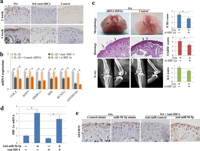
Fig. 5. HIF-2α regulates the effects of SDC-4-specific antibodies and miR-96-5p on murine osteoarthritis and chondrocyte hypertrophy.
a OA mice were injected with 10 μl of SDC-4-specific antibody or an equivalent volume of saline into the knee joint cavity. Immunohistochemistry of HIF-2α expression was performed in cartilage tissue at 2 and 4 weeks post operation. b Chondrocytes were transfected with si-HIF-2α or control siRNA for 24 h, pretreated with SDC-4-specific antibody for 2 h and then stimulated with or without recombinant human IL-1β for 4 h. The expression of COL-X, SOX9, MMP-13, IHH, RUNX2, and ADAMTS5 was determined by RT-PCR analyses in chondrocytes. c OA mice were administered si-HIF-2α or control siRNA in the knee joint cavity. Morphological, histological, and radiographic analyses of the femoral condyles were performed using digital camera, safranin O staining, and X-ray at 4 weeks post operation, respectively. The cartilage lesions were graded on a scale of ICRS; the joint lesions were graded using the OARSI scoring system; and the radiographic changes were graded using the K–L scales. The dotted circles indicate wear area. The black arrows indicate chondrofibrosis and cartilage lesions. The white arrows indicate the degree of joint space narrowing. Bar = 2 mm (morphological) and 100 μm (histological). d RT-PCR analysis of HIF-2α expression in chondrocytes transfected with miR-96-5p mimics, anti-miR-96-5p, mimic-control or anti-miRNA-control, or/and SDC-4-specific antibody. e Immunohistochemistry analysis of ADAMTS5 expression in the cartilage tissue of murine osteoarthritis treated with miR-96-5p mimics, anti-miR-96-5p, or/and SDC-4-specific antibody. Bar = 50 μm.

