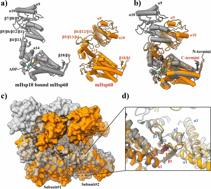
Figure 5.
mHsp10 binding stabilizes mHsp60 subunit association. (a) Ribbon diagrams of mHsp60-mHsp10 (PDB: 6MRD, grey) and mHsp60 (orange) subunits. Structural elements mentioned in text are annotated. (b) Overlay of one mHsp60-mHsp10 subunit and one mHsp60 subunit in ribbon diagram representation. (c) Overlay of mHsp60 with mHsp60-mHsp10 in surface representation. The mHsp60 heptameric ring in mHsp60-mHsp10 was superimposed with the apo mHsp60 heptamer. Only two subunits from each structure are shown. (d) Closeup on the inter-subunit interface around the β1-β18-β2-β3 sheet. The sheet acts as a pivotal point to bring the neighboring equatorial domains closer in mHsp60-mHsp10 than in mHsp60. Secondary structures are labeled in blue for one subunit and in red for the other subunit.

