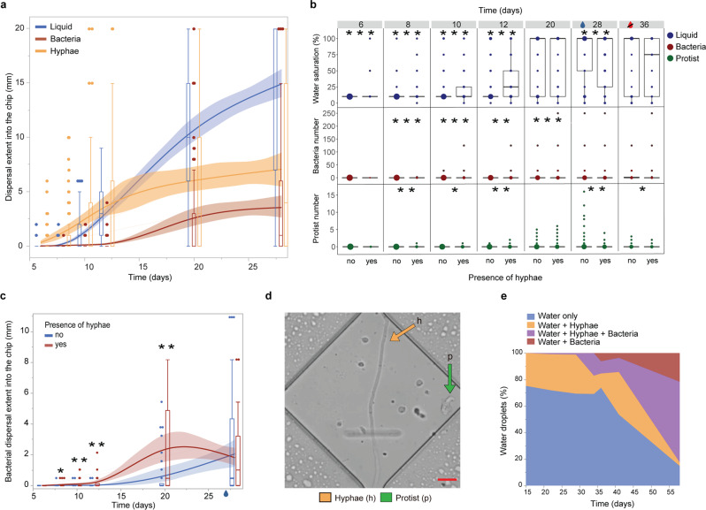
Fig. 4. Time-resolved investigation of the influence of fungal hyphae on pore wetting and microbial dispersal recorded in Expt. 3: soil incubated on initially air-filled chips in the laboratory.
a Dispersal extent of fungal hyphae, water films (liquid), and bacteria into the ‘diamond’ channels, mean and 95%-confidence interval combined with boxplots with median, quartiles and outliers, n = 3 chips × 36 channels. b Presence of water (liquid) and abundance of bacteria and protists in diamond-shape widenings over time, depending on the presence of a fungal hypha. Prior to day 28, we performed a waterlogging treatment to the inoculation soil that saturated most parts of the chips’ pore spaces, indicated by the blue droplet in b and c. Between days 29 and 36, chips were exposed to drying without any additional watering, indicated by the strikethrough red droplet in b. Boxplots with median, quartiles and outliers and the dot size indicating the number of observations, n = 3 chips × 1188 widenings. c Bacterial dispersal extent into channels with diamond-shaped widenings, depending on the presence of fungal hyphae, boxplots combined with a curve of the means and 95%-confidence interval, n = 3 chips × 36 channels. Prior to day 28, we performed a waterlogging treatment to the inoculation soil that saturated most parts of the chips’ pore spaces, indicated by the blue droplet. d Example of a diamond-shaped widening along the channels with a protist along a hypha. Scale bar = 10 µm. e Quantification of the presence of hyphae and bacteria contained within spontaneously formed water droplets within the entry system of an initially air-filled chip. Humidity of the inoculation soil was kept equal, no waterlogging or drying was applied. Stars in b and c denote statistically significant differences at *p < 0.05; **p < 0.01; ***p < 0.0001.

