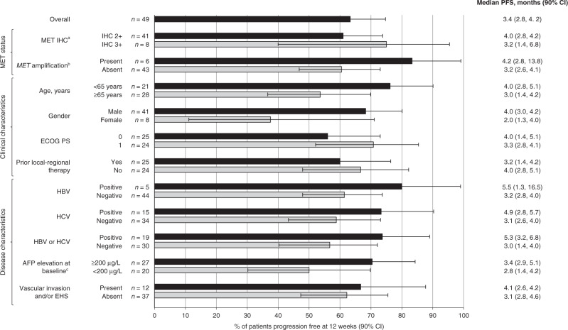
Fig. 1. Percentage of patients progression free at 12 weeks according to investigator assessment in Phase 2.
Data are shown for the overall population and patient subgroups.aModerate (2+) or strong (3+) staining intensity for MET on IHC in the majority (≥50%) of tumour cells; bMET amplification defined as MET:CEP7 ratio ≥2 or mean gene copy number ≥5; cAFP was missing for two patients (12-week PFS in these patients was 100%). AFP alpha-fetoprotein; CI confidence interval, EHS extrahepatic spread, ECOG PS Eastern Cooperative Oncology Group performance status, HBV hepatitis B virus, HCV hepatitis C virus, IHC immunohistochemistry, PFS progression-free survival.

