Figure 1.
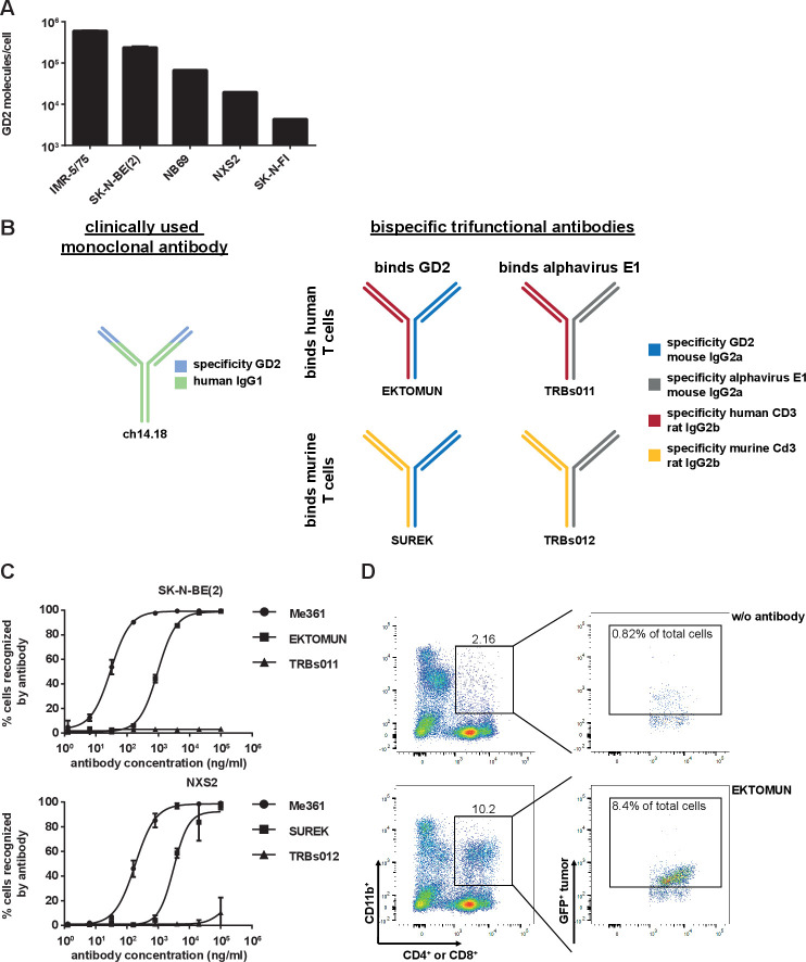
SUREK and EKTOMUN bispecific trifunctional antibodies bind to GD2 positive neuroblastoma cell lines and form effector-target cell clusters. (A) Flow cytometric quantification of GD2 molecules on the cell surface of five different neuroblastoma cell lines. (B) Schematic description of the antibodies in this study. blue: mouse IgG2a recognizing GD2; gray: mouse IgG2a recognizing the alphavirus glycoprotein E1; red: rat IgG2b recognizing human CD3; yellow: rat IgG2b recognizing mouse Cd3; green-light blue: ch14.18 a humanized chimeric antibody consisting of a human IgG1 Fc fragment attached to a murine Fab region (ligand binding domain recognizing GD2). (C) Flow cytometric binding analysis of the bispecific trifunctional antibodies SUREK, EKTOMUN, TRBs011 and TRBs012 as well as the parental GD2-directed monoclonal antibody Me361 on a human (SK-N-BE(2)) and a murine (NXS2) neuroblastoma cell line. (D) Effector-target cell cluster formation is visualized by representative dot plots showing triple+ cell clusters. Triple+ events are positive for CD4 or CD8 (T cells), CD11b (monocytic accessory immune cells) and GFP (neuroblastoma cells).

